5.1 Periods of Prenatal Development
The Human Development Teaching & Learning Group
Learning Objectives
- Differentiate between development during the germinal, embryonic, and fetal periods
- Explain the process of brain development during the prenatal period
You may be familiar with the three trimesters of pregnancy, three trimesters consisting of three months each to equal the entire pregnancy of nine months. People who have recently given birth frequently refer to how they felt during their first, second, or third trimester, and this is easy to do, as the trimesters generally describe the experience of the gestational parent during these three temporally equal intervals of pregnancy. When we look at the development of the organism from zygote to fetus to newly born infant, we also examine three periods of development. However, these periods are not equal in length and they do not align with the gestational parent’s experience of the three trimesters. Actually, a full term pregnancy is considered to be 40 weeks long (which would actually be equivalent to 10, not nine, months!), and two of the three periods of development occur within the first trimester. Let’s take a look at some of the changes that take place during each of the three periods of prenatal development: the germinal period, the embryonic period, and the fetal period.
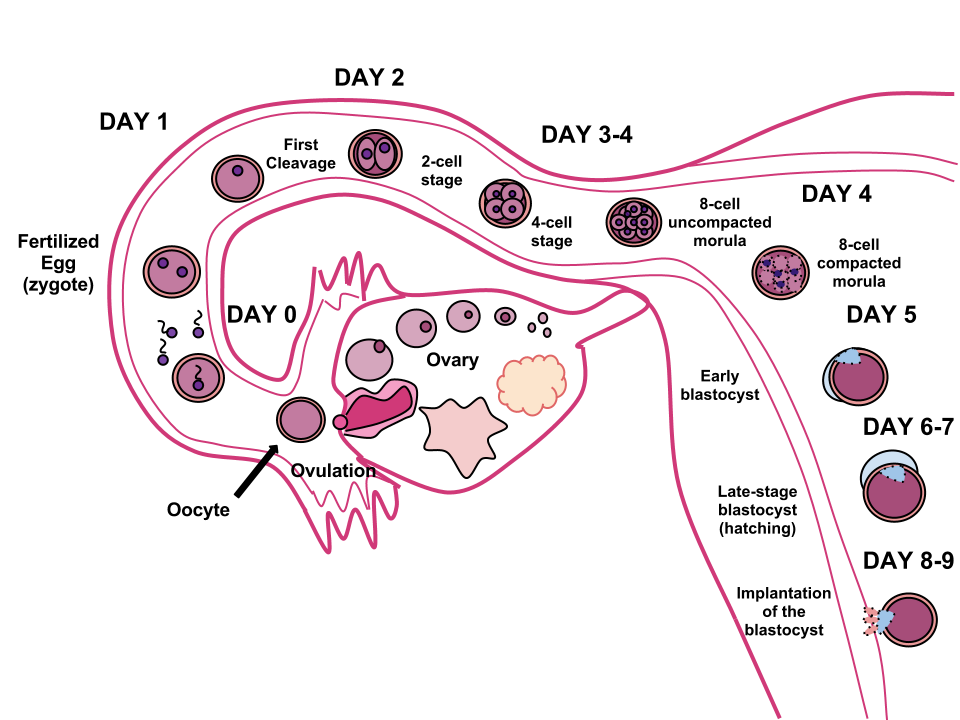
The Germinal Period (Weeks 1-2)

The germinal period (about 14 days in length) lasts from conception to implantation of the fertilized egg in the lining of the uterus (see Figure 5.3). At ejaculation millions of sperm are released into the vagina, but only a few reach the egg and typically only one fertilizes the egg. Once a single sperm has entered the wall of the egg, the wall becomes hard and prevents other sperm from entering. After the sperm has entered the egg, the tail of the sperm breaks off and the head of the sperm, containing the genetic information from the father, unites with the nucleus of the egg. The egg is typically fertilized in the top section of the fallopian tube and continues its journey to the uterus. As a result, a new cell is formed. This cell, containing the combined genetic information from both parents, is referred to as a zygote.
During this time, the organism begins cell division through mitosis. After five days of mitosis there is a group of 100 cells, which is now called a blastocyst. The blastocyst consists of both an inner and outer group of cells. The inner group of cells, or embryonic disk will become the embryo, while the outer group of cells, or trophoblast, becomes the support system which nourishes the developing organism. This stage ends when the blastocyst fully implants into the uterine wall (U.S. National Library of Medicine, 2015). Approximately 50-75% of blastocysts do not implant in the uterine wall (Betts et al., 2019).
Mitosis is a fragile process and fewer than half of all zygotes survive beyond the first two weeks (Hall, 2004). Some of the reasons for this include the egg and sperm do not join properly, meaning that their genetic material does not combine; there is too little or damaged genetic material; the zygote does not replicate; or the blastocyst does not implant into the uterine wall. The failure rate is higher for in vitro conceptions. Figure 5.3 illustrates the journey of the ova from its release to its fertilization, cell duplication, and implantation into the uterine lining.
Infertility
Infertility is the inability to conceive a child or carry a child to birth, and is usually defined when an individual has not had a successful pregnancy after one year of trying to conceive. About 75 percent of causes of infertility can be identified; these include diseases, such as sexually transmitted diseases that can cause scarring of the reproductive tubes in males or females, or developmental problems frequently related to abnormal hormone levels in one of the individuals. Inadequate nutrition, especially starvation, can delay menstruation. Stress can also lead to infertility. Short-term stress can affect hormone levels, while long-term stress can delay puberty and cause less frequent menstrual cycles. Other factors that affect fertility include toxins (such as cadmium), tobacco smoking, marijuana use, gonadal injuries, and aging.
If infertility is identified, several assisted reproductive technologies (ART) are available to aid conception. A common type of ART is in vitro fertilization (IVF) where an egg and sperm are combined outside the body and then placed in the uterus. This technique was first successfully performed in mice by Anne McLaren and John Biggers, whose work is credited as among the most significant in reproductive medicine. Eggs are obtained after extensive hormonal treatments that prepare mature eggs for fertilization and prepare the uterus for implantation of the fertilized egg. Sperm are obtained, combined with the eggs, and supported through several cell divisions to ensure viability of the zygotes. When the embryos have reached the eight-cell stage, one or more is implanted into the gestational parent’s or carrier’s uterus. If fertilization is not accomplished by simple IVF, a procedure that injects the sperm into an egg can be used. This is called intracytoplasmic sperm injection (ICSI) and is shown in Figure 5.4. IVF procedures produce a surplus of fertilized eggs and embryos that can be frozen and stored for future use. The procedures can also result in multiple births.
Watch It
The following video explains how In Vitro Fertilization (IVF) works as a solution for couples who cannot conceive naturally (e.g., some older couples and some queer couples).
The Embryonic Period (Weeks 3-8)

Starting the third week the blastocyst has implanted in the uterine wall. Upon implantation this multi-cellular organism is called an embryo. Now blood vessels grow, forming the placenta. The placenta is a structure connected to the uterus that provides nourishment and oxygen from the mother to the developing embryo via the umbilical cord. During this period, cells continue to differentiate. Growth during prenatal development occurs in two major directions: from head to tail called cephalocaudal development and from the midline outward referred to as proximodistal development. This means that those structures nearest the head develop before those nearest the feet and those structures nearest the torso develop before those away from the center of the body (such as hands and fingers). The head develops in the fourth week and the precursor to the heart begins to pulse. In the early stages of the embryonic period, gills and a tail are apparent. However, by the end of this stage they disappear and the organism takes on a more human appearance. Some organisms fail during the embryonic period, usually due to gross chromosomal abnormalities. As in the case of the germinal period, often the gestational parent does not yet know that they are pregnant.
It is during this stage that the major structures of the body are taking form making the embryonic period the time when the organism is most vulnerable to the greatest amount of damage if exposed to harmful substances. Gestational parents are not often aware of the risks they introduce to the developing embryo during this time. The embryo is approximately 1 inch in length and weighs about 8 grams at the end of eight weeks (Betts et al., 2019). The embryo can move and respond to touch at this time.
The Fetal Period (Weeks 9-40)

From the ninth week until birth, the organism is referred to as a fetus. During this stage, the major structures are continuing to develop. By the 12th week, the fetus has all its body parts including external genitalia. In the following weeks, the fetus will develop hair, nails, teeth and the excretory and digestive systems will continue to develop. The fetus is about 3 inches long and weighs about 28 grams.
During the 4th – 6th months, the eyes become more sensitive to light and hearing develops. The respiratory system continues to develop, and reflexes such as sucking, swallowing and hiccupping, develop during the 5th month. Cycles of sleep and wakefulness are present at this time as well. The first chance of survival outside the womb, known as the age of viability is reached at about 24 weeks (Morgan et al., 2008). The majority of the neurons in the brain have developed by 24 weeks, although they are still rudimentary, and the glial or nurse cells that support neurons continue to grow. At 24 weeks the fetus can feel pain (Royal College of Obstetricians and Gynecologists, 1997). By the time the fetus reaches this point, it weighs up to 1.4 pounds. The hearing has developed, so the fetus can respond to sounds. The internal organs, such as the lungs, heart, stomach, and intestines, have formed enough that a fetus born prematurely at this point has a chance to survive outside of the gestational parent’s womb.
Between the 7th – 9th months, the fetus is primarily preparing for birth. It is exercising its muscles and its lungs begin to expand and contract. The fetus gains about 5 pounds and 7 inches during this last trimester of pregnancy, and during the 8th month a layer of fat develops under the skin. This layer of fat serves as insulation and helps the baby regulate body temperature after birth.
At around 36 weeks the fetus is almost ready for birth. It weighs about 6 pounds and is about 18.5 inches long. By week 37 all of the fetus’s organ systems are developed enough that it could survive outside the gestational parent’s uterus without many of the risks associated with premature birth. The fetus continues to gain weight and grow in length until approximately 40 weeks. By then the fetus has very little room to move around and birth becomes imminent. The progression through the stages is shown in Figure 5.6.

Watch It
This video explains many of the developmental milestones and changes that happen during each month of development for the embryo and fetus.
Prenatal Brain Development
Prenatal brain development begins in the third gestational week with the differentiation of stem cells, which are capable of producing all the different cells that make up the brain (Stiles & Jernigan, 2010). The location of these stem cells in the embryo is referred to as the neural plate. By the end of the third week, two ridges appear along the neural plate first forming the neural groove and then the neural tube. The open region in the center of the neural tube forms the brain’s ventricles and spinal canal. By the end of the embryonic period, or week eight, the neural tube has further differentiated into the forebrain, midbrain, and hindbrain.
Brain development during the fetal period involves neuron production, migration, and differentiation. From the early fetal period until midgestation, most of the 85 billion neurons have been generated and many have already migrated to their brain positions. Neurogenesis, or the formation of neurons, is largely completed after five months of gestation. One exception is in the hippocampus, which continues to develop neurons throughout life. Neurons that form the neocortex, or the layer of cells that lie on the surface of the brain, migrate to their location in an orderly way. Neural migration is mostly completed in the cerebral cortex by 24 weeks (Poduri & Volpe, 2018).
Once in position, neurons begin to produce dendrites and axons that begin to form the neural networks responsible for information processing. Regions of the brain that contain the cell bodies are referred to as the gray matter because they look gray in appearance. The axons that form the neural pathways make up the white matter because they are covered in myelin, a fatty substance that is white in appearance. Myelin aids in both the insulation and efficiency of neural transmission. Although cell differentiation is complete at birth, the growth of dendrites, axons, and synapses continue for years.
Try It
References (Click to expand)
Betts, J. G., DeSaix, P., Johnson, E., Johnson, J. E., Korol, O., Kruse, D. H., Poe, B., Wise, J. A., & Young, K. A. (2019). Anatomy and physiology (OpenStax). Houston, TX: Rice University.
Hall, D. (2004). Meiotic drive and sex chromosome cycling. Evolution, 58(5), 925-931. Health Resources and Services Administration. (2015). HIV screening for pregnant women. Retrieved from http://www.hrsa.gov/quality/toolbox/measures/hivpregnantwomen/index.html
Morgan, M.A., Goldenberg, R.L., & Schulkin, J. (2008) Obstetrician-gynecologists’ practices regarding preterm birth at the limit of viability. The Journal of Maternal-Fetal and Neonatal Medicine, 21, 115-121.
Poduri, A., & Volpe, J. (2018). Volpe’s neurology of the newborn (6th edition). Amsterdam, Netherlands: Elsevier.
Royal College of Obstetricians and Gynecologists. (1997). Fetal awareness: Review of research and recommendations. Retrieved from https://www.rcog.org.uk/en/guidelines-research-services/guidelines/fetal-awareness—review-of-research-and-recommendations-for-practice/
Stiles, J. & Jernigan, T. L. (2010). The basics of brain development. Neuropsychology Review, 20(4), 327-348. doi: 10.1007/s11065-010-9148-4
United States National Library of Medicine. (2015). Fetal development. Retrieved from https://www.nlm.nih.gov/medlineplus/ency/article/002398.htm
Licenses & Attributions (Click to expand)
CC Licensed Content
- Human Pregnancy and Birth. Authored by: Mary Ann Clark, Matthew Douglas, Jung Choi. Provided by: OpenStax. Located at: https://openstax.org/books/biology-2e/pages/43-5-human-pregnancy-and-birth. License: CC BY: Attribution
- “Lifespan Development: A Psychological Perspective, Second Edition” by Martha Lally and Suzanne Valentine-French is licensed under a CC-BY-NC-SA-3.0
- Prenatal Development. Authored by: Julie Lazzara for Lumen Learning. Provided by: Lumen Learning. Located at: https://courses.lumenlearning.com/wm-lifespandevelopment/chapter/prenatal-development/. License: CC BY: Attribution
- Psyc 200 Lifespan Psychology. Authored by: Laura Overstreet. Located at: http://opencourselibrary.org/econ-201/. License: CC BY: Attribution
Media Attributions
- How in vitro fertilization (IVF) works by TED is licensed CC BY–NC–ND 4.0
- Prenatal Development content and fetal stages image. Provided by: Lumen Learning. Located at: https://courses.lumenlearning.com/waymaker-psychology/chapter/stages-of-development/. License: CC BY: Attribution
- Fetus image. Authored by: Jacopo Werther. Provided by: Wikimedia. Located at: https://commons.wikimedia.org/wiki/File:Human_fetus_10_weeks_with_amniotic_sac_-_therapeutic_abortion.jpg. License: CC BY-SA: Attribution-ShareAlike
- Sperm-egg © Unknown is licensed under a Public Domain license
- Human_Fertilization © Ttrue12 is licensed under a CC BY-SA (Attribution ShareAlike) license
- Human_Embryo_-_Approximately_8_weeks_estimated_gestational_age © Lunar Caustic is licensed under a CC BY (Attribution) license
- prenataldev © Martha Lally and Suzanne Valentine-French is licensed under a CC BY-NC-SA (Attribution NonCommercial ShareAlike) license
All Rights Reserved Content
- Prenatal Development: What Babies Learn Inside the Womb. Authored by: sprouts. Located at: https://www.youtube.com/watch?time_continue=175&v=UA-Tk9qlG9A. License: Other. License Terms: Standard YouTube License
