Ch 5: Biopsychology

Introduction to Neural Communication
Ever wonder how your brain actually works? What exactly is going on inside of your small, wrinkly mass while you read this text? In this section, you’ll learn about the basics of neural communication in the brain, which is the brain’s way of sending messages to and from different regions in order to relay critical information about your body and its surroundings.
Glia and neurons are the two cell types that make up the nervous system. While glia generally play supporting roles, the communication between neurons is fundamental to all of the functions associated with the nervous system. Neuronal communication is made possible by the neuron’s specialized structures, like the soma, dendrites, axons, terminal buttons, and synaptic vesicles.
Neuronal communication is an electrochemical event. The dendrites contain receptors for neurotransmitters released by nearby neurons. If the signals received from other neurons are sufficiently strong, an action potential will travel down the length of the axon to the terminal buttons, resulting in the release of neurotransmitters into the synapse.
Different neurotransmitters are associated with different functions. Often, psychological disorders involve imbalances in a given neurotransmitter system. Therefore, psychotropic drugs are prescribed in an attempt to bring the neurotransmitters back into balance. Drugs can act either as agonists or as antagonists for a given neurotransmitter system.
Neural Communication
Learning Objectives
- Explain the role and function of the basic structures of a neuron
- Describe how neurons communicate with each other
- Explain how drugs act as agonists or antagonists for a given neurotransmitter system
Neurons
Psychologists striving to understand the human mind may study the nervous system. Learning how the cells and organs (like the brain) function, help us understand the biological basis behind human psychology. The nervous system is composed of two basic cell types: glial cells (also known as glia) and neurons. Glial cells, which outnumber neurons ten to one, are traditionally thought to play a supportive role to neurons, both physically and metabolically. Glial cells provide scaffolding on which the nervous system is built, help neurons line up closely with each other to allow neuronal communication, provide insulation to neurons, transport nutrients and waste products, and mediate immune responses. Neurons, on the other hand, serve as interconnected information processors that are essential for all of the tasks of the nervous system. This section briefly describes the structure and function of neurons.
Neuron Structure
Neurons are the central building blocks of the nervous system, 100 billion strong at birth. Like all cells, neurons consist of several different parts, each serving a specialized function. A neuron’s outer surface is made up of a semipermeable membrane. This membrane allows smaller molecules and molecules without an electrical charge to pass through it, while stopping larger or highly charged molecules.
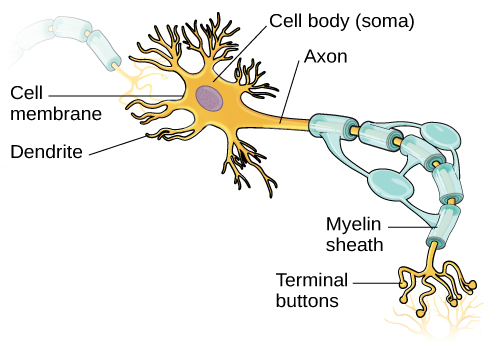
The nucleus of the neuron is located in the soma, or cell body. The soma has branching extensions known as dendrites. The neuron is a small information processor, and dendrites serve as input sites where signals are received from other neurons. These signals are transmitted electrically across the soma and down a major extension from the soma known as the axon, which ends at multiple terminal buttons. The terminal buttons contain synaptic vesicles that house neurotransmitters, the chemical messengers of the nervous system.
Axons range in length from a fraction of an inch to several feet. In some axons, glial cells form a fatty substance known as the myelin sheath, which coats the axon and acts as an insulator, increasing the speed at which the signal travels. The myelin sheath is crucial for the normal operation of the neurons within the nervous system: the loss of the insulation it provides can be detrimental to normal function. To understand how this works, let’s consider an example. Multiple sclerosis (MS), an autoimmune disorder, involves a large-scale loss of the myelin sheath on axons throughout the nervous system. The resulting interference in the electrical signal prevents the quick transmittal of information by neurons and can lead to a number of symptoms, such as dizziness, fatigue, loss of motor control, and sexual dysfunction. While some treatments may help to modify the course of the disease and manage certain symptoms, there is currently no known cure for multiple sclerosis.
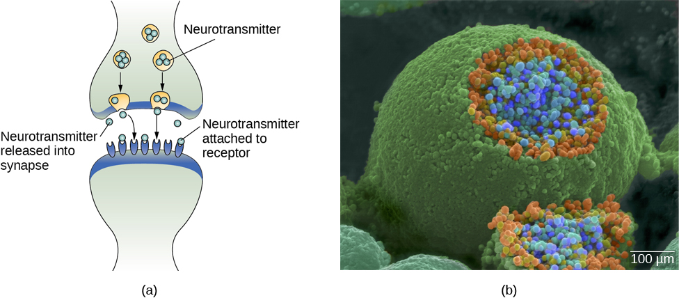
In healthy individuals, the neuronal signal moves rapidly down the axon to the terminal buttons, where synaptic vesicles release neurotransmitters into the synapse. The synapse is a very small space between two neurons and is an important site where communication between neurons occurs. Once neurotransmitters are released into the synapse, they travel across the small space and bind with corresponding receptors on the dendrite of an adjacent neuron. Receptors proteins on the cell surface where neurotransmitters attach, vary in shape, with different shapes “matching” different neurotransmitters.
Watch It
This video shows the structure and physiology of a neuron.
You can view the transcript for “2-Minute Neuroscience: The Neuron” here (opens in new window).
How does a neurotransmitter “know” which receptor to bind to? The neurotransmitter and the receptor have what is referred to as a lock-and-key relationship—specific neurotransmitters fit specific receptors similar to how a key fits a lock. The neurotransmitter binds to any receptor that it fits.
Try It h5p id=”68″]
How Neurons Communicate
Now that we have learned about the basic structures of the neuron and the role that these structures play in neuronal communication, let’s take a closer look at the signal itself—how it moves through the neuron and then jumps to the next neuron, where the process is repeated.
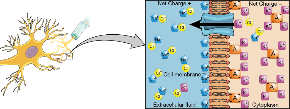
We begin at the neuronal membrane. The neuron exists in a fluid environment—it is surrounded by extracellular fluid and contains intracellular fluid (i.e., cytoplasm). The neuronal membrane keeps these two fluids separate—a critical role because the electrical signal that passes through the neuron depends on the intra- and extracellular fluids being electrically different. This difference in charge across the membrane, called the membrane potential, provides energy for the signal.
The electrical charge of the fluids is caused by charged molecules (ions) dissolved in the fluid. The semipermeable nature of the neuronal membrane somewhat restricts the movement of these charged molecules, and, as a result, some of the charged particles tend to become more concentrated either inside or outside the cell.
Between signals, the neuron membrane’s potential is held in a state of readiness, called the resting potential. Like a rubber band stretched out and waiting to spring into action, ions line up on either side of the cell membrane, ready to rush across the membrane when the neuron goes active and the membrane opens its gates (i.e., a sodium-potassium pump that allows movement of ions across the membrane). Ions in high-concentration areas are ready to move to low-concentration areas, and positive ions are ready to move to areas with a negative charge.
In the resting state, sodium (Na+) is at higher concentrations outside the cell, so it will tend to move into the cell. Potassium (K+), on the other hand, is more concentrated inside the cell, and will tend to move out of the cell (Figure 4). In addition, the inside of the cell is slightly negatively charged compared to the outside. This provides an additional force on sodium, causing it to move into the cell.
From this resting potential state, the neuron receives a signal and its state changes abruptly (Figure 5). When a neuron receives signals at the dendrites—due to neurotransmitters from an adjacent neuron binding to its receptors—small pores, or gates, open on the neuronal membrane, allowing Na+ ions, propelled by both charge and concentration differences, to move into the cell. With this influx of positive ions, the internal charge of the cell becomes more positive. If that charge reaches a certain level, called the threshold of excitation, the neuron becomes active and the action potential begins. This process of when the cell’s charge becomes positive, or less negative, is called depolarization.
Many additional pores open, causing a massive influx of Na+ ions and a huge positive spike in the membrane potential, the peak action potential. At the peak of the spike, the sodium gates close and the potassium gates open. As positively charged potassium ions leave, the cell quickly begins repolarization. At first, it hyperpolarizes, becoming slightly more negative than the resting potential, and then it levels off, returning to the resting potential.
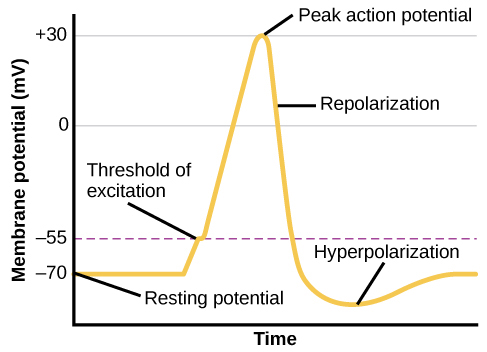
This positive spike constitutes the action potential: the electrical signal that typically moves from the cell body down the axon to the axon terminals. The electrical signal moves down the axon like a wave; at each point, some of the sodium ions that enter the cell diffuse to the next section of the axon, raising the charge past the threshold of excitation and triggering a new influx of sodium ions. The action potential moves all the way down the axon to the terminal buttons.
Watch It
The process of neural communication is explained in the following video.
You can view the transcript for “Lights, Camera, Action Potentials!” here (opens in new window).
The action potential is an all-or-none phenomenon. In simple terms, this means that an incoming signal from another neuron is either sufficient or insufficient to reach the threshold of excitation. There is no in-between, and there is no turning off an action potential once it starts. Think of it like sending an email or a text message. You can think about sending it all you want, but the message is not sent until you hit the send button. Furthermore, once you send the message, there is no stopping it.
Because it is all or none, the action potential is recreated, or propagated, at its full strength at every point along the axon. Much like the lit fuse of a firecracker, it does not fade away as it travels down the axon. It is this all-or-none property that explains the fact that your brain perceives an injury to a distant body part like your toe as equally painful as one to your nose.
As noted earlier, when the action potential arrives at the terminal button, the synaptic vesicles release their neurotransmitters into the synapse The neurotransmitters travel across the synapse and bind to receptors on the dendrites of the adjacent neuron, and the process repeats itself in the new neuron (assuming the signal is sufficiently strong to trigger an action potential). Once the signal is delivered, excess neurotransmitters in the synapse drift away, are broken down into inactive fragments, or are reabsorbed in a process known as reuptake. Reuptake involves the neurotransmitter being pumped back into the neuron that released it, in order to clear the synapse (Figure 6). Clearing the synapse serves both to provide a clear “on” and “off” state between signals and to regulate the production of neurotransmitter (full synaptic vesicles provide signals that no additional neurotransmitters need to be produced).
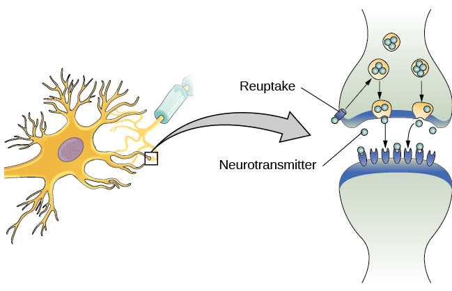
Neuronal communication is often referred to as an electrochemical event. The movement of the action potential down the length of the axon is an electrical event, and movement of the neurotransmitter across the synaptic space represents the chemical portion of the process.
Try It
Watch It
Watch the following video to see how neurons communicate within the body.
You can view the transcript for “How do nerves work? – Elliot Krane” here (opens in new window).
Neurotransmitters and Drugs
There are several different types of neurotransmitters released by different neurons, and we can speak in broad terms about the kinds of functions associated with different neurotransmitters (Table 1). Much of what psychologists know about the functions of neurotransmitters comes from research on the effects of drugs in psychological disorders. Psychologists who take a biological perspective and focus on the physiological causes of behavior assert that psychological disorders like depression and schizophrenia are associated with imbalances in one or more neurotransmitter systems. In this perspective, psychotropic medications can help improve the symptoms associated with these disorders. Psychotropic medications are drugs that treat psychiatric symptoms by restoring neurotransmitter balance.
| Neurotransmitter | Involved in | Potential Effect on Behavior |
|---|---|---|
| Acetylcholine | Muscle action, memory | Increased arousal, enhanced cognition |
| Beta-endorphin | Pain, pleasure | Decreased anxiety, decreased tension |
| Dopamine | Mood, sleep, learning | Increased pleasure, suppressed appetite |
| Gamma-aminobutyric acid (GABA) | Brain function, sleep | Decreased anxiety, decreased tension |
| Glutamate | Memory, learning | Increased learning, enhanced memory |
| Norepinephrine | Heart, intestines, alertness | Increased arousal, suppressed appetite |
| Serotonin | Mood, sleep | Modulated mood, suppressed appetite |
Psychoactive drugs can act as agonists or antagonists for a given neurotransmitter system. Agonists are chemicals that mimic a neurotransmitter at the receptor site and, thus, strengthen its effects. An antagonist, on the other hand, blocks or impedes the normal activity of a neurotransmitter at the receptor. Agonist and antagonist drugs are prescribed to correct the specific neurotransmitter imbalances underlying a person’s condition. For example, Parkinson’s disease, a progressive nervous system disorder, is associated with low levels of dopamine. Therefore dopamine agonists, which mimic the effects of dopamine by binding to dopamine receptors, are one treatment strategy.
Certain symptoms of schizophrenia are associated with overactive dopamine neurotransmission. The antipsychotics used to treat these symptoms are antagonists for dopamine—they block dopamine’s effects by binding its receptors without activating them. Thus, they prevent dopamine released by one neuron from signaling information to adjacent neurons.
In contrast to agonists and antagonists, which both operate by binding to receptor sites, reuptake inhibitors prevent unused neurotransmitters from being transported back to the neuron. This leaves more neurotransmitters in the synapse for a longer time, increasing its effects. Depression, which has been consistently linked with reduced serotonin levels, is commonly treated with selective serotonin reuptake inhibitors (SSRIs). By preventing reuptake, SSRIs strengthen the effect of serotonin, giving it more time to interact with serotonin receptors on dendrites. Common SSRIs on the market today include Prozac, Paxil, and Zoloft. The drug LSD is structurally very similar to serotonin, and it affects the same neurons and receptors as serotonin. Psychotropic drugs are not instant solutions for people suffering from psychological disorders. Often, an individual must take a drug for several weeks before seeing improvement, and many psychoactive drugs have significant negative side effects. Furthermore, individuals vary dramatically in how they respond to the drugs. To improve chances for success, it is not uncommon for people receiving pharmacotherapy to undergo psychological and/or behavioral therapies as well. Some research suggests that combining drug therapy with other forms of therapy tends to be more effective than any one treatment alone (for one such example, see March et al., 2007).
Try It
Watch It
Review the process of neural communication in the following CrashCourse psychology video:
You can view the transcript for “The Chemical Mind: Crash Course Psychology #3” here (opens in new window).
Try It
The Nervous System

In this section, you’ll learn about the basics of the central nervous system, which consists of the brain and spinal cord, as well as the peripheral nervous system. The peripheral nervous system is comprised of the somatic and autonomic nervous systems. The somatic nervous system transmits sensory and motor signals to and from the central nervous system. The autonomic nervous system controls the function of our organs and glands, and can be divided into the sympathetic and parasympathetic divisions. Sympathetic activation prepares us for fight or flight, while parasympathetic activation is associated with normal functioning under relaxed conditions. Got all that? We’ll review each of these systems in the coming pages.
The Nervous System
Learning Objectives
- Describe the difference between the central and peripheral nervous systems and the somatic and autonomic nervous systems
- Differentiate between the sympathetic and parasympathetic divisions of the autonomic nervous system
Parts of the Nervous System
The nervous system can be divided into two major subdivisions: the central nervous system (CNS) and the peripheral nervous system (PNS), shown in Figure 7. The CNS is comprised of the brain and spinal cord; the PNS connects the CNS to the rest of the body. In this section, we focus on the peripheral nervous system; later, we look at the brain and spinal cord.

Peripheral Nervous System
The peripheral nervous system is made up of thick bundles of axons, called nerves, carrying messages back and forth between the CNS and the muscles, organs, and senses in the periphery of the body (i.e., everything outside the CNS). The PNS has two major subdivisions: the somatic nervous system and the autonomic nervous system.
The somatic nervous system is associated with activities traditionally thought of as conscious or voluntary. It is involved in the relay of sensory and motor information to and from the CNS; therefore, it consists of motor neurons and sensory neurons. Motor neurons, carrying instructions from the CNS to the muscles, are efferent fibers (efferent means “moving away from”). Sensory neurons, carrying sensory information to the CNS, are afferent fibers (afferent means “moving toward”). Each nerve is basically a two-way superhighway, containing thousands of axons, both efferent and afferent.
The autonomic nervous system controls our internal organs and glands and is generally considered to be outside the realm of voluntary control. It can be further subdivided into the sympathetic and parasympathetic divisions (Figure 8). The sympathetic nervous system is involved in preparing the body for stress-related activities; the parasympathetic nervous system is associated with returning the body to routine, day-to-day operations. The two systems have complementary functions, operating in tandem to maintain the body’s homeostasis. Homeostasisis a state of equilibrium, in which biological conditions (such as body temperature) are maintained at optimal levels.

The sympathetic nervous system is activated when we are faced with stressful or high-arousal situations. The activity of this system was adaptive for our ancestors, increasing their chances of survival. Imagine, for example, that one of our early ancestors, out hunting small game, suddenly disturbs a large bear with her cubs. At that moment, his body undergoes a series of changes—a direct function of sympathetic activation—preparing him to face the threat. His pupils dilate, his heart rate and blood pressure increase, his bladder relaxes, his liver releases glucose, and adrenaline surges into his bloodstream. This constellation of physiological changes, known as the fight or flight response, allows the body access to energy reserves and heightened sensory capacity so that it might fight off a threat or run away to safety.
While it is clear that such a response would be critical for survival for our ancestors, who lived in a world full of real physical threats, many of the high-arousal situations we face in the modern world are more psychological in nature. For example, think about how you feel when you have to stand up and give a presentation in front of a roomful of people, or right before taking a big test. You are in no real physical danger in those situations, and yet you have evolved to respond to any perceived threat with the fight or flight response. This kind of response is not nearly as adaptive in the modern world; in fact, we suffer negative health consequences when faced constantly with psychological threats that we can neither fight nor flee. Recent research suggests that an increase in susceptibility to heart disease (Chandola, Brunner, & Marmot, 2006) and impaired function of the immune system (Glaser & Kiecolt-Glaser, 2005) are among the many negative consequences of persistent and repeated exposure to stressful situations.
Once the threat has been resolved, the parasympathetic nervous system takes over and returns bodily functions to a relaxed state. Our hunter’s heart rate and blood pressure return to normal, his pupils constrict, he regains control of his bladder, and the liver begins to store glucose in the form of glycogen for future use. These processes are associated with activation of the parasympathetic nervous system.
Try It
Think It Over
Try It
Parts of the Brain
 In this section, you’ll learn about the specific parts of the brain and their roles and functions. While this is not an anatomy class, you’ll see how important it is to understand the parts of the brain and what they do so that we can understand mental processes and behavior.
Watch It
Watch this CrashCourse Psychology video for an overview on the brain and the interesting topics we’ll cover:
Part of the Brain
Learning Objectives
- Explain the two hemispheres of the brain, lateralization and plasticity
- Identify the location and function of the lobes of the brain
- Identify and describe the role of the parts of the limbic system, the midbrain, and hindbrain
- Describe the types of techniques available to clinicians and researchers to image or scan the brain
Brain Hemispheres
The central nervous system (CNS), consists of the brain and the spinal cord.
Brain
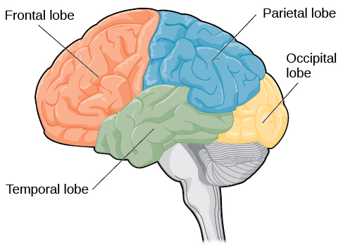
The brain is a remarkably complex organ comprised of billions of interconnected neurons and glia. It is a bilateral, or two-sided, structure that can be separated into distinct lobes. Each lobe is associated with certain types of functions, but, ultimately, all of the areas of the brain interact with one another to provide the foundation for our thoughts and behaviors.
Spinal Cord
It can be said that the spinal cord is what connects the brain to the outside world. Because of it, the brain can act. The spinal cord is like a relay station, but a very smart one. It not only routes messages to and from the brain, but it also has its own system of automatic processes, called reflexes.
The top of the spinal cord merges with the brain stem, where the basic processes of life are controlled, such as breathing and digestion. In the opposite direction, the spinal cord ends just below the ribs—contrary to what we might expect, it does not extend all the way to the base of the spine.
The spinal cord is functionally organized in 30 segments, corresponding with the vertebrae. Each segment is connected to a specific part of the body through the peripheral nervous system. Nerves branch out from the spine at each vertebra. Sensory nerves bring messages in; motor nerves send messages out to the muscles and organs. Messages travel to and from the brain through every segment.
Some sensory messages are immediately acted on by the spinal cord, without any input from the brain. Withdrawal from heat and knee jerk are two examples. When a sensory message meets certain parameters, the spinal cord initiates an automatic reflex. The signal passes from the sensory nerve to a simple processing center, which initiates a motor command. Seconds are saved, because messages don’t have to go the brain, be processed, and get sent back. In matters of survival, the spinal reflexes allow the body to react extraordinarily fast.
The spinal cord is protected by bony vertebrae and cushioned in cerebrospinal fluid, but injuries still occur. When the spinal cord is damaged in a particular segment, all lower segments are cut off from the brain, causing paralysis. Therefore, the lower on the spine damage is, the fewer functions an injured individual loses.
Two Hemispheres
The surface of the brain, known as the cerebral cortex, is very uneven, characterized by a distinctive pattern of folds or bumps, known as gyri (singular: gyrus), and grooves, known as sulci (singular: sulcus), shown in Figure 9. These gyri and sulci form important landmarks that allow us to separate the brain into functional centers. The most prominent sulcus, known as the longitudinal fissure, is the deep groove that separates the brain into two halves or hemispheres: the left hemisphere and the right hemisphere.

There is evidence of some specialization of function—referred to as lateralization—in each hemisphere, mainly regarding differences in language ability. Beyond that, however, the differences that have been found have been minor (this means that it is a myth that a person is either left-brained dominant or right-brained dominant).[1] What we do know is that the left hemisphere controls the right half of the body, and the right hemisphere controls the left half of the body.
The two hemispheres are connected by a thick band of neural fibers known as the corpus callosum, consisting of about 200 million axons. The corpus callosum allows the two hemispheres to communicate with each other and allows for information being processed on one side of the brain to be shared with the other side.
Normally, we are not aware of the different roles that our two hemispheres play in day-to-day functions, but there are people who come to know the capabilities and functions of their two hemispheres quite well. In some cases of severe epilepsy, doctors elect to sever the corpus callosum as a means of controlling the spread of seizures (Figure 10). While this is an effective treatment option, it results in individuals who have split brains. After surgery, these split-brain patients show a variety of interesting behaviors. For instance, a split-brain patient is unable to name a picture that is shown in the patient’s left visual field because the information is only available in the largely nonverbal right hemisphere. However, they are able to recreate the picture with their left hand, which is also controlled by the right hemisphere. When the more verbal left hemisphere sees the picture that the hand drew, the patient is able to name it (assuming the left hemisphere can interpret what was drawn by the left hand).

Much of what we know about the functions of different areas of the brain comes from studying changes in the behavior and ability of individuals who have suffered damage to the brain. For example, researchers study the behavioral changes caused by strokes to learn about the functions of specific brain areas. A stroke, caused by an interruption of blood flow to a region in the brain, causes a loss of brain function in the affected region. The damage can be in a small area, and, if it is, this gives researchers the opportunity to link any resulting behavioral changes to a specific area. The types of deficits displayed after a stroke will be largely dependent on where in the brain the damage occurred.
Consider Theona, an intelligent, self-sufficient woman, who is 62 years old. Recently, she suffered a stroke in the front portion of her right hemisphere. As a result, she has great difficulty moving her left leg. (As you learned earlier, the right hemisphere controls the left side of the body; also, the brain’s main motor centers are located at the front of the head, in the frontal lobe.) Theona has also experienced behavioral changes. For example, while in the produce section of the grocery store, she sometimes eats grapes, strawberries, and apples directly from their bins before paying for them. This behavior—which would have been very embarrassing to her before the stroke—is consistent with damage in another region in the frontal lobe—the prefrontal cortex, which is associated with judgment, reasoning, and impulse control.
Watch It
Watch this video to see an incredible example of the challenges facing a split-brain patient shortly following the surgery to sever her corpus callosum.
You can view the transcript for “Split Brain mpeg1video” here (opens in new window).
Watch this second video about another patient who underwent a dramatic surgery to prevent her seizures. You’ll learn more about the brain’s ability to change, adapt, and reorganize itself, also known as brain plasticity.
You can view the transcript for “Brain Plasticity – the story of Jody” here (opens in new window).
Try It
Lobes of the Brain
Forebrain Structures

Lobes of the Brain
The four lobes of the brain are the frontal, parietal, temporal, and occipital lobes (Figure 12). The frontal lobe is located in the forward part of the brain, extending back to a fissure known as the central sulcus. The frontal lobe is involved in reasoning, motor control, emotion, and language. It contains the motor cortex, which is involved in planning and coordinating movement; the prefrontal cortex, which is responsible for higher-level cognitive functioning; and Broca’s area, which is essential for language production.
People who suffer damage to Broca’s area have great difficulty producing language of any form. For example, Padma was an electrical engineer who was socially active and a caring, involved mother. About twenty years ago, she was in a car accident and suffered damage to her Broca’s area. She completely lost the ability to speak and form any kind of meaningful language. There is nothing wrong with her mouth or her vocal cords, but she is unable to produce words. She can follow directions but can’t respond verbally, and she can read but no longer write. She can do routine tasks like running to the market to buy milk, but she could not communicate verbally if a situation called for it.
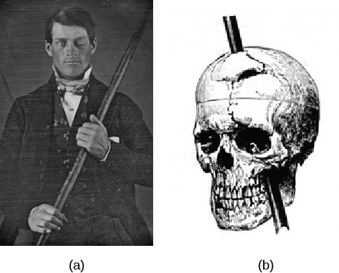
Probably the most famous case of frontal lobe damage is that of a man by the name of Phineas Gage. On September 13, 1848, Gage (age 25) was working as a railroad foreman in Vermont. He and his crew were using an iron rod to tamp explosives down into a blasting hole to remove rock along the railway’s path. Unfortunately, the iron rod created a spark and caused the rod to explode out of the blasting hole, into Gage’s face, and through his skull (Figure 13). Although lying in a pool of his own blood with brain matter emerging from his head, Gage was conscious and able to get up, walk, and speak. But in the months following his accident, people noticed that his personality had changed. Many of his friends described him as no longer being himself. Before the accident, it was said that Gage was a well-mannered, soft-spoken man, but he began to behave in odd and inappropriate ways after the accident. Such changes in personality would be consistent with loss of impulse control—a frontal lobe function.
Beyond the damage to the frontal lobe itself, subsequent investigations into the rod’s path also identified probable damage to pathways between the frontal lobe and other brain structures, including the limbic system. With connections between the planning functions of the frontal lobe and the emotional processes of the limbic system severed, Gage had difficulty controlling his emotional impulses.
However, there is some evidence suggesting that the dramatic changes in Gage’s personality were exaggerated and embellished. Gage’s case occurred in the midst of a 19th century debate over localization—regarding whether certain areas of the brain are associated with particular functions. On the basis of extremely limited information about Gage, the extent of his injury, and his life before and after the accident, scientists tended to find support for their own views, on whichever side of the debate they fell (Macmillan, 1999).
Watch It
Watch this clip about Phineas Gage to learn more about his accident and injury.
You can view the transcript for “Phineas Gage (LEGO Stop-Motion Video)” (opens in new window).
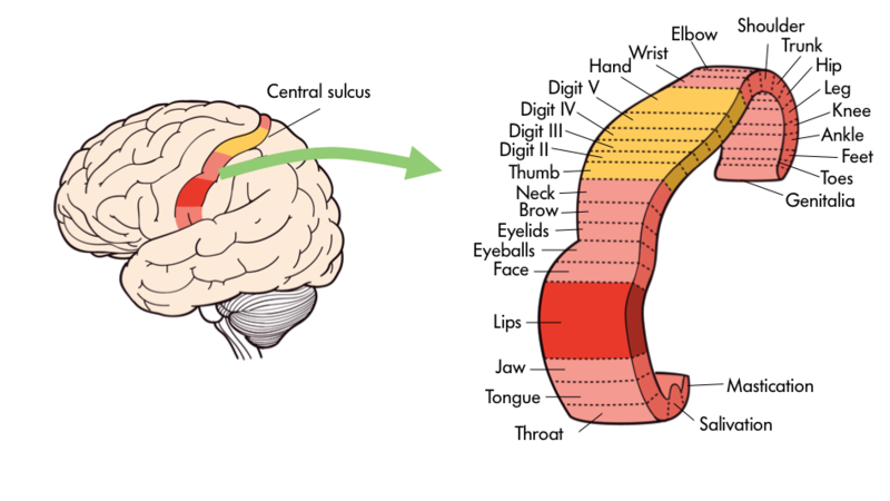
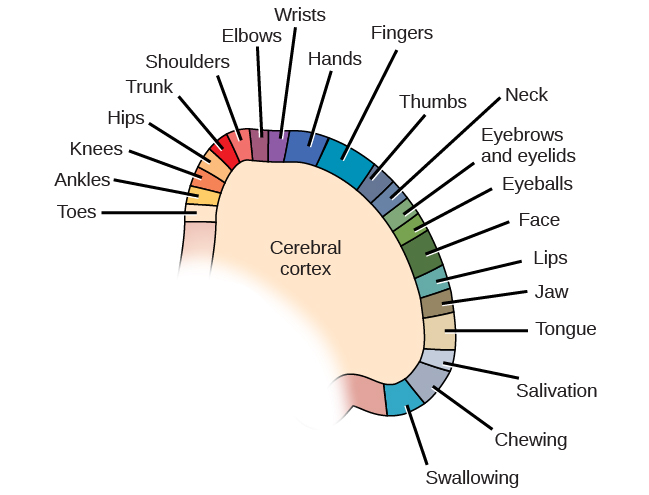
One particularly fascinating area in the frontal lobe is called the “primary motor cortex”. This strip running along the side of the brain is in charge of voluntary movements like waving goodbye, wiggling your eyebrows, and kissing. It is an excellent example of the way that the various regions of the brain are highly specialized. Interestingly, each of our various body parts has a unique portion of the primary motor cortex devoted to it. Each individual finger has about as much dedicated brain space as your entire leg. Your lips, in turn, require about as much dedicated brain processing as all of your fingers and your hand combined!
Because the cerebral cortex in general, and the frontal lobe in particular, are associated with such sophisticated functions as planning and being self-aware they are often thought of as a higher, less primal portion of the brain. Indeed, other animals such as rats and kangaroos while they do have frontal regions of their brain do not have the same level of development in the cerebral cortices. The closer an animal is to humans on the evolutionary tree—think chimpanzees and gorillas, the more developed is this portion of their brain.
The brain’s parietal lobe is located immediately behind the frontal lobe, and is involved in processing information from the body’s senses. It contains the somatosensory cortex, which is essential for processing sensory information from across the body, such as touch, temperature, and pain. The somatosensory cortex is organized topographically, which means that spatial relationships that exist in the body are maintained on the surface of the somatosensory cortex. For example, the portion of the cortex that processes sensory information from the hand is adjacent to the portion that processes information from the wrist.
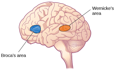
The temporal lobe is located on the side of the head (temporal means “near the temples”), and is associated with hearing, memory, emotion, and some aspects of language. The auditory cortex, the main area responsible for processing auditory information, is located within the temporal lobe. Wernicke’s area, important for speech comprehension, is also located here. Whereas individuals with damage to Broca’s area have difficulty producing language, those with damage to Wernicke’s area can produce sensible language, but they are unable to understand it.
The occipital lobe is located at the very back of the brain, and contains the primary visual cortex, which is responsible for interpreting incoming visual information. The occipital cortex is organized retinotopically, which means there is a close relationship between the position of an object in a person’s visual field and the position of that object’s representation on the cortex. You will learn much more about how visual information is processed in the occipital lobe when you study sensation and perception.
Try It
Think It Over
Consider the following advice from Joseph LeDoux, a professor of neuroscience and psychology at New York University, as you learn about the specific parts of the brain:
Be suspicious of any statement that says a brain area is a center responsible for some function. The notion of functions being products of brain areas or centers is left over from the days when most evidence about brain function was based on the effects of brain lesions localized to specific areas. Today, we think of functions as products of systems rather than of areas. Neurons in areas contribute because they are part of a system. The amygdala, for example, contributes to threat detection because it is part of a threat detection system. And just because the amygdala contributes to threat detection does not mean that threat detection is the only function to which it contributes. Amygdala neurons, for example, are also components of systems that process the significance of stimuli related to eating, drinking, sex, and addictive drugs.
Try It
The Limbic System and Other Brain Areas
Areas of the Forebrain

The[pb_glossary id=”2515″] limbic system[/pb_glossary] is involved in processing both emotion and memory. Interestingly, the sense of smell projects directly to the limbic system; therefore, not surprisingly, smell can evoke emotional responses in ways that other sensory modalities cannot. The limbic system is made up of a number of different structures, but three of the most important are the hippocampus, the amygdala, and the hypothalamus (Figure 18). The hippocampus is an essential structure for learning and memory. The amygdala is involved in our experience of emotion and in tying emotional meaning to our memories. The hypothalamus regulates a number of homeostatic processes, including the regulation of body temperature, appetite, and blood pressure. The hypothalamus also serves as an interface between the nervous system and the endocrine system and in the regulation of sexual motivation and behavior.

Case Study: Henry Molaison (H.M.)
Try It
Link to Learning
Midbrain and Hindbrain Structures
The midbrain is comprised of structures located deep within the brain, between the forebrain and the hindbrain. The reticular formation is centered in the midbrain, but it actually extends up into the forebrain and down into the hindbrain. The reticular formation is important in regulating the sleep/wake cycle, arousal, alertness, and motor activity.
The substantia nigra (Latin for “black substance”) and the ventral tegmental area (VTA) are also located in the midbrain (Figure 19). Both regions contain cell bodies that produce the neurotransmitter dopamine, and both are critical for movement. Degeneration of the substantia nigra and VTA is involved in Parkinson’s disease. In addition, these structures are involved in mood, reward, and addiction (Berridge & Robinson, 1998; Gardner, 2011; George, Le Moal, & Koob, 2012).

The hindbrain is located at the back of the head and looks like an extension of the spinal cord. It contains the medulla, pons, and cerebellum (Figure 20). The medulla controls the automatic processes of the autonomic nervous system, such as breathing, blood pressure, and heart rate. The word pons literally means “bridge,” and as the name suggests, the pons serves to connect the brain and spinal cord. It also is involved in regulating brain activity during sleep. The medulla, pons, and midbrain together are known as the brainstem.

The cerebellum (Latin for “little brain”) receives messages from muscles, tendons, joints, and structures in our ear to control balance, coordination, movement, and motor skills. The cerebellum is also thought to be an important area for processing some types of memories. In particular, procedural memory, or memory involved in learning and remembering how to perform tasks, is thought to be associated with the cerebellum. Recall that H. M. was unable to form new explicit memories, but he could learn new tasks. This is likely due to the fact that H. M.’s cerebellum remained intact.
Link to Learning
For a fun recap of the parts of the brain, watch the following short clip from the old cartoon, Pinky and the Brain:
You can view the transcript for “pinky and the brain-brainstem” here (opens in new window).
Dig Deeper: Brain Dead and on Life Support
What would you do if your spouse or loved one was declared brain dead but his or her body was being kept alive by medical equipment? Whose decision should it be to remove a feeding tube? Should medical care costs be a factor?
On February 25, 1990, a Florida woman named Terri Schiavo went into cardiac arrest, apparently triggered by a bulimic episode. She was eventually revived, but her brain had been deprived of oxygen for a long time. Brain scans indicated that there was no activity in her cerebral cortex, and she suffered from severe and permanent cerebral atrophy. Basically, Schiavo was in a vegetative state. Medical professionals determined that she would never again be able to move, talk, or respond in any way. To remain alive, she required a feeding tube, and there was no chance that her situation would ever improve.
On occasion, Schiavo’s eyes would move, and sometimes she would groan. Despite the doctors’ insistence to the contrary, her parents believed that these were signs that she was trying to communicate with them.
After 12 years, Schiavo’s husband argued that his wife would not have wanted to be kept alive with no feelings, sensations, or brain activity. Her parents, however, were very much against removing her feeding tube. Eventually, the case made its way to the courts, both in the state of Florida and at the federal level. By 2005, the courts found in favor of Schiavo’s husband, and the feeding tube was removed on March 18, 2005. Schiavo died 13 days later.
Why did Schiavo’s eyes sometimes move, and why did she groan? Although the parts of her brain that control thought, voluntary movement, and feeling were completely damaged, her brainstem was still intact. Her medulla and pons maintained her breathing and caused involuntary movements of her eyes and the occasional groans. Over the 15-year period that she was on a feeding tube, Schiavo’s medical costs may have topped $7 million (Arnst, 2003).
These questions were brought to popular conscience 25 years ago in the case of Terri Schiavo, and they persist today. In 2013, a 13-year-old girl who suffered complications after tonsil surgery was declared brain dead. There was a battle between her family, who wanted her to remain on life support, and the hospital’s policies regarding persons declared brain dead. In another complicated 2013–14 case in Texas, a pregnant EMT professional declared brain dead was kept alive for weeks, despite her spouse’s directives, which were based on her wishes should this situation arise. In this case, state laws designed to protect an unborn fetus came into consideration until doctors determined the fetus unviable.
Decisions surrounding the medical response to patients declared brain dead are complex. What do you think about these issues?
Try It
Think It Over
Try It
Brain Imaging
Techniques Involving Radiation

A computerized tomography (CT) scan involves taking a number of x-rays of a particular section of a person’s body or brain (Figure 21). The x-rays pass through tissues of different densities at different rates, allowing a computer to construct an overall image of the area of the body being scanned. A CT scan is often used to determine whether someone has a tumor, or significant brain atrophy.

Positron emission tomography (PET) scans create pictures of the living, active brain (Figure 22). An individual receiving a PET scan drinks or is injected with a mildly radioactive substance, called a tracer. Once in the bloodstream, the amount of tracer in any given region of the brain can be monitored. As brain areas become more active, more blood flows to that area. A computer monitors the movement of the tracer and creates a rough map of active and inactive areas of the brain during a given behavior. PET scans show little detail, are unable to pinpoint events precisely in time, and require that the brain be exposed to radiation; therefore, this technique has been replaced by the fMRI as an alternative diagnostic tool. However, combined with CT, PET technology is still being used in certain contexts. For example, CT/PET scans allow better imaging of the activity of neurotransmitter receptors and open new avenues in schizophrenia research. In this hybrid CT/PET technology, CT contributes clear images of brain structures, while PET shows the brain’s activity.

Techniques Involving Magnetic Fields
In magnetic resonance imaging (MRI), a person is placed inside a machine that generates a strong magnetic field. The magnetic field causes the hydrogen atoms in the body’s cells to move. When the magnetic field is turned off, the hydrogen atoms emit electromagnetic signals as they return to their original positions. Tissues of different densities give off different signals, which a computer interprets and displays on a monitor.
Functional magnetic resonance imaging (fMRI) operates on the same principles, but it shows changes in brain activity over time by tracking blood flow and oxygen levels. The fMRI provides more detailed images of the brain’s structure, as well as better accuracy in time, than is possible in PET scans (Figure 23). With their high level of detail, MRI and fMRI are often used to compare the brains of healthy individuals to the brains of individuals diagnosed with psychological disorders. This comparison helps determine what structural and functional differences exist between these populations.
Link to Learning
Techniques Involving Electrical Activity
In some situations, it is helpful to gain an understanding of the overall activity of a person’s brain, without needing information on the actual location of the activity. Electroencephalography (EEG) serves this purpose by providing a measure of a brain’s electrical activity. An array of electrodes is placed around a person’s head (Figure 24). The signals received by the electrodes result in a printout of the electrical activity of his or her brain, or brainwaves, showing both the frequency (number of waves per second) and amplitude (height) of the recorded brainwaves, with an accuracy within milliseconds. Such information is especially helpful to researchers studying sleep patterns among individuals with sleep disorders.

Try It
Nature and Nurture
 How do we become who we are? Traditionally, people’s answers have placed them in one of two camps: nature or nurture. The one says genes determine an individual while the other claims the environment is the linchpin for development. Since the 16th century, when the terms “nature” and “nurture” first came into use, many people have spent ample time debating which is more important, but these discussions have more often led to ideological cul-de-sacs rather than pinnacles of insight.
New research into epigenetics—the science of how the environment influences genetic expression—is changing the conversation. As psychologist David S. Moore explains in his newest book, The Developing Genome, this burgeoning field reveals that what counts is not what genes you have so much as what your genes are doing. And what your genes are doing is influenced by the ever-changing environment they’re in. Factors like stress, nutrition, and exposure to toxins all play a role in how genes are expressed—essentially which genes are turned on or off. Unlike the static conception of nature or nurture, epigenetic research demonstrates how genes and environments continuously interact to produce characteristics throughout a lifetime.
Nature and Nurture
Learning Objectives
- Describe epigenetics and examine how gene-environment interactions are critical for expression of physical and psychological characteristics
Gene-Environment Interactions
Genes do not exist in a vacuum. Although we are all biological organisms, we also exist in an environment that is incredibly important in determining not only when and how our genes express themselves, but also in what combination. Each of us represents a unique interaction between our genetic makeup and our environment; range of reaction is one way to describe this interaction. Range of reaction asserts that our genes set the boundaries within which we can operate, and our environment interacts with the genes to determine where in that range we will fall. For example, if an individual’s genetic makeup predisposes her to high levels of intellectual potential and she is reared in a rich, stimulating environment, then she will be more likely to achieve her full potential than if she were raised under conditions of significant deprivation. According to the concept of range of reaction, genes set definite limits on potential, and environment determines how much of that potential is achieved.
Another perspective on the interaction between genes and the environment is the concept of genetic environmental correlation. Stated simply, our genes influence our environment, and our environment influences the expression of our genes (Figure 31). Not only do our genes and environment interact, as in range of reaction, but they also influence one another bidirectionally. For example, the child of an NBA player would probably be exposed to basketball from an early age. Such exposure might allow the child to realize his or her full genetic, athletic potential. Thus, the parents’ genes, which the child shares, influence the child’s environment, and that environment, in turn, is well suited to support the child’s genetic potential.

In another approach to gene-environment interactions, the field of epigenetics looks beyond the genotype itself and studies how the same genotype can be expressed in different ways. In other words, researchers study how the same genotype can lead to very different phenotypes. As mentioned earlier, gene expression is often influenced by environmental context in ways that are not entirely obvious. For instance, identical twins share the same genetic information. Identical twins develop from a single fertilized egg that split, so the genetic material is exactly the same in each; in contrast, fraternal twins develop from two different eggs fertilized by different sperm, so the genetic material varies as with non-twin siblings). But even with identical genes, there remains an incredible amount of variability in how gene expression can unfold over the course of each twin’s life. Sometimes, one twin will develop a disease and the other will not. In one example, Tiffany, an identical twin, died from cancer at age 7, but her twin, now 19 years old, has never had cancer. Although these individuals share an identical genotype, their phenotypes differ as a result of how that genetic information is expressed over time. The epigenetic perspective is very different from range of reaction, because here the genotype is not fixed and limited.
Watch It
You can view the transcript for “Introducing Epigenetics – Intro to Psychology” here (opens in new window).
Visit this website on genetics for an engaging video primer on the epigenetics of twin studies.

Genes affect more than our physical characteristics. Indeed, scientists have found genetic linkages to a number of behavioral characteristics, ranging from basic personality traits to sexual orientation to spirituality (for examples, see Mustanski et al., 2005; Comings, Gonzales, Saucier, Johnson, & MacMurray, 2000). Genes are also associated with temperament and a number of psychological disorders, such as depression and schizophrenia. So while it is true that genes provide the biological blueprints for our cells, tissues, organs, and body, they also have significant impact on our experiences and our behaviors.Let’s look at the following findings regarding schizophrenia in light of our three views of gene-environment interactions. Which view do you think best explains this evidence?In a study of people who were given up for adoption, adoptees whose biological mothers had schizophrenia and who had been raised in a disturbed family environment were much more likely to develop schizophrenia or another psychotic disorder than were any of the other groups in the study:
- Of adoptees whose biological mothers had schizophrenia (high genetic risk) and who were raised in disturbed family environments, 36.8% were likely to develop schizophrenia.
- Of adoptees whose biological mothers had schizophrenia (high genetic risk) and who were raised in healthy family environments, 5.8% were likely to develop schizophrenia.
- Of adoptees with a low genetic risk (whose mothers did not have schizophrenia) and who were raised in disturbed family environments, 5.3% were likely to develop schizophrenia.
- Of adoptees with a low genetic risk (whose mothers did not have schizophrenia) and who were raised in healthy family environments, 4.8% were likely to develop schizophrenia (Tienari et al., 2004).
The study shows that adoptees with high genetic risk were especially likely to develop schizophrenia only if they were raised in disturbed home environments. This research lends credibility to the notion that both genetic vulnerability and environmental stress are necessary for schizophrenia to develop, and that genes alone do not tell the full tale.
Dig Deeper: Parental Investment and programming of stress responses in Offspring
The most comprehensive study to date of variations in parental investment and epigenetic inheritance in mammals is that of the maternally transmitted responses to stress in rats. In rat pups, maternal nurturing (licking and grooming) during the first week of life is associated with long-term programming of individual differences in stress responsiveness, emotionality, cognitive performance, and reproductive behavior (Caldji et al., 1998; Francis, Diorio, Liu, & Meaney, 1999; Liu et al., 1997; Myers, Brunelli, Shair, Squire, & Hofer, 1989; Stern, 1997). In adulthood, the offspring of mothers that exhibit increased levels of pup licking and grooming over the first week of life show increased expression of the glucocorticoid receptor in the hippocampus (a brain structure associated with stress responsivity as well as learning and memory) and a lower hormonal response to stress compared with adult animals reared by low licking and grooming mothers (Francis et al., 1999; Liu et al., 1997). Moreover, rat pups that received low levels of maternal licking and grooming during the first week of life showed decreased histone acetylation and increased DNA methylation of a neuron-specific promoter of the glucocorticoid receptor gene (Weaver et al., 2004). The expression of this gene is then reduced, the number of glucocorticoid receptors in the brain is decreased, and the animals show a higher hormonal response to stress throughout their life.
The effects of maternal care on stress hormone responses and behavior in the offspring can be eliminated in adulthood by pharmacological treatment (HDAC inhibitor trichostatin A, TSA) or dietary amino acid supplementation (methyl donor L-methionine), treatments that influence histone acetylation, DNA methylation, and expression of the glucocorticoid receptor gene (Weaver et al., 2004;Weaver et al., 2005). This series of experiments shows that histone acetylation and DNA methylation of the glucocorticoid receptor gene promoter is a necessary link in the process leading to the long-term physiological and behavioral sequelae of poor maternal care. This points to a possible molecular target for treatments that may reverse or ameliorate the traces of childhood maltreatment.
Several studies have attempted to determine to what extent the findings from model animals are transferable to humans. Examination of post-mortem brain tissue from healthy human subjects found that the human equivalent of the glucocorticoid receptor gene promoter (NR3C1 exon 1F promoter) is also unique to the individual (Turner, Pelascini, Macedo, & Muller, 2008). A similar study examining newborns showed that methylation of the glucocorticoid receptor gene promoter maybe an early epigenetic marker of maternal mood and risk of increased hormonal responses to stress in infants 3 months of age (Oberlander et al., 2008).
Although further studies are required to examine the functional consequence of this DNA methylation, these findings are consistent with our studies in the neonate and adult offspring of low licking and grooming mothers that show increased DNA methylation of the promoter of the glucocorticoid receptor gene, decreased glucocorticoid receptor gene expression, and increased hormonal responses to stress (Weaver et al., 2004).
Examination of brain tissue from suicide victims found that the human glucocorticoid receptor gene promoter is also more methylated in the brains of individuals who had experienced maltreatment during childhood (McGowan et al., 2009). Examination of blood samples from adult patients with bipolar disorder, who also retrospectively reported on their experiences of childhood abuse and neglect, found that the degree of DNA methylation of the human glucocorticoid receptor gene promoter was strongly positively related to the reported experience of childhood maltreatment decades earlier.
Watch this video to see another example of how diet can alter the phenotype of genetically identical mice.
Try It
Biopsychology
Learning Objectives
In this chapter, you learned to
- identify the basic structures of a neuron, the function of each structure, and how messages travel through the neuron
- describe the role of the nervous system and endocrine systems
- identify and describe the parts of the brain
- explain how nature, nurture, and epigenetics influence personality and behavior
Read the following abstract from Lane Beckes, James A. Coan, and Karen Hasselmo’s 2012 study, “Familiarity promotes the blurring of self and other in the neural representation of threat.”
Neurobiological investigations of empathy often support an embodied simulation account. Using functional magnetic resonance imaging (fMRI), we monitored statistical associations between brain activations indicating self-focused threat to those indicating threats to a familiar friend or an unfamiliar stranger. Results in regions such as the anterior insula, putamen and supramarginal gyrus indicate that self-focused threat activations are robustly correlated with friend-focused threat activations but not stranger-focused threat activations. These results suggest that one of the defining features of human social bonding may be increasing levels of overlap between neural representations of self and other. This article presents a novel and important methodological approach to fMRI empathy studies, which informs how differences in brain activation can be detected in such studies and how covariate approaches can provide novel and important information regarding the brain and empathy.
Did you recognize any of the concepts discussed in this module? This study used fMRI to examine the brain activation of people as they looked at cues and received, or were threatened with receiving, mild electric shocks while holding hands with either a friend or a stranger. The results showed the expected response—brain activation in the anterior insula, putamen, and supramarginal gyrus when a person was threatened with a shock. What was remarkable, however, was that people showed nearly the same brain activation when a friend was threatened with the shock, but not a stranger. This provides insight into studies on empathy, and the idea that the concept of “self” can expand to include others as well.
As you can see, there is a limitless amount of information that could be studied on the brain. Neuroscience is a relatively new field, but the more research that is done, the more it appears that much of human behavior and mental processes—the key interests for psychological study—are intimately intertwined with activity in the brain. Understanding the brain is important no matter what type of psychology you will be involved with, because its effects permeate all human behavior.
Watch It
The more we learn about the brain and its functioning, the better able we are to work towards repairing the brain or mimicking its capabilities. These advances in research lead to medical discoveries and breakthroughs, such as the one explained in the following video:
You can view the transcript for “The nerve bypass: how to move a paralysed hand” here (opens in new window).
Chapter References (Click to expand)
Arnst, C. (2003, November). Commentary: Getting rational about health-care rationing. Bloomberg Businessweek Magazine. Retrieved from http://www.businessweek.com/stories/2003-11-16/commentary-getting-rational-about-health-care-rationing
Berridge, K. C., & Robinson, T. E. (1998). What is the role of dopamine in reward: Hedonic impact, reward learning, or incentive salience? Brain Research Reviews, 28, 309–369.
Bouchard, T. J., Lykken, D. T., McGue, M., Segal, N. L., & Tellegen, A. (1990). Sources of human psychological differences: The Minnesota Study of Twins Reared Apart. Science, 250, 223–228.
Caldji, C., Tannenbaum, B., Sharma, S., Francis, D., Plotsky, P. M., & Meaney, M. J. (1998). Maternal care during infancy regulates the development of neural systems mediating the expression of fearfulness in the rat. Proceedings of the National Academy of Sciences U S A, 95(9), 5335–5340.
Chandola, T., Brunner, E., & Marmot, M. (2006). Chronic stress at work and the metabolic syndrome: A prospective study. BMJ, 332, 521–524.
Comings, D. E., Gonzales, N., Saucier, G., Johnson, J. P., & MacMurray, J. P. (2000). The DRD4 gene and the spiritual transcendence scale of the character temperament index. Psychiatric Genetics, 10, 185–189.
Confer, J. C., Easton, J. A., Fleischman, D. S., Goetz, C. D., Lewis, D. M. G, Perilloux, C., & Buss, D. M. (2010). Evolutionary psychology: Controversies, questions, prospects, and limitations. American Psychologist, 65, 110–126.
Francis, D., Diorio, J., Liu, D., & Meaney, M. J. (1999). Nongenomic transmission across generations of maternal behavior and stress responses in the rat. Science, 286(5442), 1155–1158.
Gaines, C. (2013, August). An A-Rod suspension would save the Yankees as much as $37.5 million in 2014 alone. Business Insider. Retrieved from http://www.businessinsider.com/an-a-rod-suspension-would-save-the-yankees-as-much-as-375-million-in-2014-2013-8
Gardner, E. L. (2011). Addiction and brain reward and antireward pathways. Advances in Psychosomatic Medicine, 30, 22–60.
George, O., Le Moal, M., & Koob, G. F. (2012). Allostasis and addiction: Role of the dopamine and corticotropin-releasing factor systems. Physiology & Behavior, 106, 58–64.
Glaser, R., & Kiecolt-Glaser, J. K. (2005). Stress-induced immune dysfunction: Implications for health. Nature Reviews Immunology, 5, 243–251.
Gong, L., Parikh, S., Rosenthal, P. J., & Greenhouse, B. (2013). Biochemical and immunological mechanisms by which sickle cell trait protects against malaria. Malaria Journal. Advance online publication. doi:10.1186/1475-2875-12-317
Hardt, O., Einarsson, E. Ö., & Nader, K. (2010). A bridge over troubled water: Reconsolidation as a link between cognitive and neuroscientific memory research traditions. Annual Review of Psychology, 61, 141–167.
Liu, D., Diorio, J., Tannenbaum, B., Caldji, C., Francis, D., Freedman, A., . . . Meaney, M. J. (1997). Maternal care, hippocampal glucocorticoid receptors, and hypothalamic- pituitary-adrenal responses to stress [see comments]. Science, 277(5332), 1659–1662.
Macmillan, M. (1999). The Phineas Gage Information Page. Retrieved from http://www.uakron.edu/gage
March, J. S., Silva, S., Petrycki, S., Curry, J., Wells, K., Fairbank, J., … Severe, J. (2007). The treatment for adolescents with depression study (TADS): Long-term effectiveness and safety outcomes. Arch Gen Psychiatry, 64, 1132–1143.
McGowan, P. O., Sasaki, A., D’Alessio, A. C., Dymov, S., Labonte, B., Szyf, M., . . . Meaney, M. J. (2009). Epigenetic regulation of the glucocorticoid receptor in human brain associates with childhood abuse. Nature Neuroscience, 12(3), 342–348. doi: nn.2270 [pii]10.1038/nn.2270
McGue, M., & Lykken, D. T. (1992). Genetic influence on risk of divorce. Psychological Science, 3(6), 368–373.
Mustanski, B. S., DuPree, M. G., Nievergelt, C. M., Bocklandt, S., Schork, N. J., & Hamer, D. H. (2005). A genome wide scan of male sexual orientation. Human Genetics, 116, 272–278.
Myers, M. M., Brunelli, S. A., Shair, H. N., Squire, J. M., & Hofer, M. A. (1989). Relationships between maternal behavior of SHR and WKY dams and adult blood pressures of cross-fostered F1 pups. Developmental Psychobiology, 22(1), 55–67.
Nielsen, J. A., Zielinsk,i B. A., Ferguson ,M. A., Lainhart J. E., & Anderson, J. S. (2013). An Evaluation of the Left-Brain vs. Right-Brain Hypothesis with Resting State Functional Connectivity Magnetic Resonance Imaging. PLoS ONE 8(8): e71275. https://doi.org/10.1371/journal.pone.0071275
Oberlander, T. F., Weinberg, J., Papsdorf, M., Grunau, R., Misri, S., & Devlin, A. M. (2008). Prenatal exposure to maternal depression, neonatal methylation of human glucocorticoid receptor gene (NR3C1) and infant cortisol stress responses. Epigenetics, 3(2), 97–106. doi: 6034 [pii]
Plomin, R., Corley, R., DeFries, J. C., & Fulker, D. W. (1990). Individual differences in television viewing in early childhood: Nature as well as nurture. Psychological Science, 1(6), 371–377.
Plomin, R., DeFries, J. C., Knopik, V. S., & Neiderhiser, J. M. (2012). Behavioral genetics. New York, NY: Worth Publishers.
Stern, J. M. (1997). Offspring-induced nurturance: animal-human parallels. Developmental Psychobiololgy, 31(1), 19–37.
Squire, L. R. (2009). The legacy of patient H. M. for neuroscience. Neuron, 61, 6–9.
Tienari, P., Wynne, L. C., Sorri, A., et al. (2004). Genotype–environment interaction in schizophrenia spectrum disorder: long-term follow-up study of Finnish adoptees. British Journal of Psychiatry, 184, 216–222.
Turner, J. D., Pelascini, L. P., Macedo, J. A., & Muller, C. P. (2008). Highly individual methylation patterns of alternative glucocorticoid receptor promoters suggest individualized epigenetic regulatory mechanisms. Nucleic Acids Research, 36(22), 7207–7218. doi: gkn897 [pii] 10.1093/nar/gkn897
Weaver, I. C., Cervoni, N., Champagne, F. A., D’Alessio, A. C., Sharma, S., Seckl, J. R., . . . Meaney, M. J. (2004). Epigenetic programming by maternal behavior. Nature Neuroscience, 7(8), 847–854. doi: 10.1038/nn1276
Weaver, I. C., Champagne, F. A., Brown, S. E., Dymov, S., Sharma, S., Meaney, M. J., & Szyf, M. (2005). Reversal of maternal programming of stress responses in adult offspring through methyl supplementation: altering epigenetic marking later in life. Journal of Neuroscience, 25(47), 11045–11054. doi: 10.1523/JNEUROSCI.3652-05.2005
Licenses and Attributions (Click to expand)
CC licensed content, Original
- Biopsychology. Authored by: Karenna Malavanti Provided by: PressBooks. License: CC BY-NC-SA: Attribution-NonCommercial-ShareAlike
CC licensed content, Shared Previously
- Introduction to Neural Communication. Authored by: Lumen Learning Provided by: Lumen Learning. License: CC BY-SA: Attribution-ShareAlike Located at: https://pressbooks.online.ucf.edu/lumenpsychology/chapter/outcome-neurons/
- Neuron in tissue culture. License: CC BY-SA: Attribution-ShareAlike. Located at: https://commons.wikimedia.org/wiki/File:Neuron_in_tissue_culture.jpg.
- Cells of the Nervous System. Authored by: OpenStax College. License: CC-BY: Attribution. License Terms: Download for free at https://openstax.org/books/psychology-2e/pages/1-introduction Located at: https://openstax.org/books/psychology-2e/pages/3-2-cells-of-the-nervous-system.
- Neurons. Authored by: Lumen Learning Provided by: Lumen Learning. License: CC BY: Attribution Located at: https://pressbooks.online.ucf.edu/lumenpsychology/chapter/cells-of-the-nervous-system/
- How Neurons Communicate. Authored by: Lumen Learning Provided by: Lumen Learning. License: CC BY: Attribution Located at: https://pressbooks.online.ucf.edu/lumenpsychology/chapter/reading-neural-communication/
- Introduction to The Nervous System and the Endocrine System. Authored by: Lumen Learning Provided by: Lumen Learning. License: CC BY: Attribution Located at: https://pressbooks.online.ucf.edu/lumenpsychology/chapter/outcome-the-brain/
- Parts of the Nervous System. Authored by: OpenStax College. License: CC BY: Attribution. License Terms: Download for free at https://openstax.org/books/psychology-2e/pages/1-introduction Located at: https://openstax.org/books/psychology-2e/pages/3-3-parts-of-the-nervous-system.
- Introduction to the Parts of the Brain. Authored by: Lumen Learning Provided by: Lumen Learning. License: CC BY: Attribution Located at: https://pressbooks.online.ucf.edu/lumenpsychology/chapter/outcome-parts-of-the-brain/
- Brain Hemispheres. Authored by: Lumen Learning Provided by: Lumen Learning. License: CC BY: Attribution Located at: https://pressbooks.online.ucf.edu/lumenpsychology/chapter/the-brain-and-spinal-cord/
- The Brain and Spinal Cord. Authored by: OpenStax College. License: CC BY: Attribution. License Terms: Download for free at https://openstax.org/books/psychology-2e/pages/1-introduction/. Located at: https://openstax.org/books/psychology-2e/pages/3-4-the-brain-and-spinal-cord.
- Lobes of the Brain. Authored by: Lumen Learning Provided by: Lumen Learning. License: CC BY-NC-SA: Attribution-NonCommercial-ShareAlike Located at: https://pressbooks.online.ucf.edu/lumenpsychology/chapter/reading-parts-of-the-brain/https://pressbooks.online.ucf.edu/lumenpsychology/chapter/reading-parts-of-the-brain/
- The Amygdala Is Not The Brains Fear Center. Authored by: Joseph LeDoux. Project: The Psych Report. License: CC BY-NC-SA: Attribution-NonCommercial-ShareAlike Located at: http://thepsychreport.com/science/the-amygdala-is-not-the-brains-fear-center/.
- Motor cortex paragraphs and image. Authored by: Robert Biswas-Diener. Provided by: Portland State University. Project: The Noba Project. License: CC BY-NC-SA: Attribution-NonCommercial-ShareAlike Located at: http://nobaproject.com/modules/the-brain-and-nervous-system.
- The Limbic System and Other Brain Areas. Authored by: Lumen Learning Provided by: Lumen Learning. License: CC BY-SA: Attribution-ShareAlike Located at: https://pressbooks.online.ucf.edu/lumenpsychology/chapter/reading-the-limbic-system-and-other-brain-areas/
- Brain Imaging. Authored by: Lumen Learning Provided by: Lumen Learning. License: CC BY-SA: Attribution-ShareAlike Located at: https://pressbooks.online.ucf.edu/lumenpsychology/chapter/1993/
- Brain Review Activity. Authored by: Jessica Traylor for Lumen Learning. Provided by: Lumen Learning. License: CC BY: Attribution
- DNA image. Provided by: Wikimedia. License: CC BY-SA: Attribution-ShareAlike Located at: https://simple.wikipedia.org/wiki/DNA_methylation#/media/File:DNA_methylation.jpg.
- The End of Nature Versus Nurture. Authored by: Evan Nesterak. Project: The Psych Report. License: CC BY-NC-SA: Attribution-NonCommercial-ShareAlike. Located at: http://thepsychreport.com/books/the-end-of-nature-versus-nurture/.
- Epigenetics in Psychology (Dig Deeper activity and image of twins). Authored by: Ian Weaver. Provided by: Dalhousie University. Project: The Noba Project. License: CC BY-NC-SA: Attribution-NonCommercial-ShareAlike. Located at: http://nobaproject.com/modules/epigenetics-in-psychology?r=LDExNDY3.
- Putting It Together: Biopsychology. Authored by: Lumen Learning Provided by: Lumen Learning. License: CC BY-NC-SA: Attribution-NonCommercial-ShareAlike Located at: https://pressbooks.online.ucf.edu/lumenpsychology/chapter/putting-it-together-biopsychology/
- Familiarity promotes the blurring of self and other in the neural representation of threat. Authored by: Lane Beckes, James A. Coan, and Karen Hasselmo. Provided by: Oxford Academic. Project: Social Cognitive and Affective Neuroscience (2012) 8 (6): 670-677. License: CC BY-NC: Attribution-NonCommercial. Located at: https://academic.oup.com/scan/article/8/6/670/1611749/Familiarity-promotes-the-blurring-of-self-and.
- Psychology and the Brain. Provided by: Boundless. Project: Boundless Psychology. License: CC BY-SA: Attribution-ShareAlike. Located at: https://www.boundless.com/psychology/textbooks/boundless-psychology-textbook/biological-foundations-of-psychology-3/psychology-and-the-brain-406/studying-the-brain-149-12684/.
All Rights Reserved Content
- 2-Minute Neuroscience: The Neuron. Authored by: Neuroscientifically Challenged. License: Other. License Terms: Standard YouTube License. Located at: https://www.youtube.com/watch?v=6qS83wD29PY.
- The Chemical Mind – Crash Course Psychology #3. Authored by: Hank Green. Provided by: CrashCourse. License: Other. License Terms: Standard YouTube License Located at: https://www.youtube.com/watch?v=W4N-7AlzK7s.
- Lights, Camera, Action Potentials!. Authored by: Carleton University. License: Other. License Terms: Standard YouTube License Located at: https://youtu.be/XdCrZm_JAp0?t=1m20s.
- How do nerves work? . Authored by: Elliot Krane. Provided by: TedEd. License: Other. License Terms: Standard YouTube License Located at: https://www.youtube.com/watch?v=uU_4uA6-zcE#t=64.
- Meet Your Master: Getting to Know Your Brain – Crash Course Psychology #4. Provided by: CrashCourse. License: Other. License Terms: Standard YouTube License. Located at: https://www.youtube.com/watch?v=vHrmiy4W9C0.
- Split Brain mpeg1video. Authored by: mrsrooboy. License: Other. License Terms: Standard YouTube License Located at: https://www.youtube.com/watch?v=8C8qu8FnuAo&feature=youtu.be.
- Brain Plasticity – the story of Jody. Authored by: Streetwisdom Billy. License: Other. License Terms: Standard YouTube License Located at: https://www.youtube.com/watch?v=VaDlLD97CLM&feature=youtu.be.
- Phineas Gage (LEGO Stop-Motion Music Video). Authored by: Brad Wray. License: Other. License Terms: Standard YouTube License. Located at: https://www.youtube.com/watch?v=_nikOxNfjqs.
- pinky and the brain-brainstem. Authored by: ctdalilah. License: Other. License Terms: Standard YouTube License. Located at: https://www.youtube.com/watch?v=snO68aJTOpM.
- Introducing epigenetics – Intro to Psychology. Authored by: Udacity. License: Other. License Terms: Standard YouTube License. Located at: https://www.youtube.com/watch?v=JCZ-flJ-QxA.
- The nerve bypass: how to move a paralysed hand. Provided by: Nature. License: Other. License Terms: Standard YouTube License. Located at: https://www.youtube.com/watch?v=60fAjaRfwnU.
Public domain content
- Spinal Cord. Authored by: BruceBlaus. Provided by: Wikimedia. License: CC BY: Attribution Located at: https://en.wikipedia.org/wiki/Surfer%27s_myelopathy#/media/File:Blausen_0822_SpinalCord.png.
- brain image. Authored by: Henry Gray. Provided by: Wikimedia. License: Public Domain: No Known Copyright Located at: https://commons.wikimedia.org/wiki/File:Lobes_of_the_brain_NL.svg.
- Nielsen JA, Zielinski BA, Ferguson MA, Lainhart JE, Anderson JS (2013) An Evaluation of the Left-Brain vs. Right-Brain Hypothesis with Resting State Functional Connectivity Magnetic Resonance Imaging. PLoS ONE 8(8): e71275. https://doi.org/10.1371/journal.pone.0071275 ↵
nervous system cell that provides physical and metabolic support to neurons, including neuronal insulation and communication, and nutrient and waste transport
cells in the nervous system that act as interconnected information processors, which are essential for all of the tasks of the nervous system
cell body
branch-like extension of the soma that receives incoming signals from other neurons
major extension of the soma
axon terminal containing synaptic vesicles
storage site for neurotransmitters
chemical messenger of the nervous system
fatty substance that insulates axons
small gap between two neurons where communication occurs
protein on the cell surface where neurotransmitters attach
difference in charge across the neuronal membrane
the state of readiness of a neuron membrane’s potential between signals
level of charge in the membrane that causes the neuron to become active
process of when the cell's charge becomes positive, or less negative
neuron becoming slightly more negative than the resting potential
electrical signal that moves down the neuron’s axon
phenomenon that incoming signal from another neuron is either sufficient or insufficient to reach the threshold of excitation
neurotransmitter is pumped back into the neuron that released it
view that psychological disorders like depression and schizophrenia are associated with imbalances in one or more neurotransmitter systems
drugs that treat psychiatric symptoms by restoring neurotransmitter balance
drug that mimics or strengthens the effects of a neurotransmitter
drug that blocks or impedes the normal activity of a given neurotransmitter
connects the brain and spinal cord to the muscles, organs and senses in the periphery of the body
relays sensory and motor information to and from the CNS
controls our internal organs and glands
involved in stress-related activities and functions
associated with routine, day-to-day operations of the body
brain and spinal cord
state of equilibrium—biological conditions, such as body temperature, are maintained at optimal levels
activation of the sympathetic division of the autonomic nervous system, allowing access to energy reserves and heightened sensory capacity so that we might fight off a given threat or run away to safety
surface of the brain that is associated with our highest mental capabilities
(plural: gyri) bump or ridge on the cerebral cortex
(plural: sulci) depressions or grooves in the cerebral cortex
deep groove in the brain’s cortex
concept that each hemisphere of the brain is associated with specialized functions
left or right half of the brain
thick band of neural fibers connecting the brain’s two hemispheres
largest part of the brain, containing the cerebral cortex, the thalamus, and the limbic system, among other structures
part of the cerebral cortex involved in reasoning, motor control, emotion, and language; contains motor cortex
strip of cortex involved in planning and coordinating movement
area in the frontal lobe responsible for higher-level cognitive functioning
region in the left hemisphere that is essential for language production
part of the cerebral cortex involved in processing various sensory and perceptual information; contains the primary somatosensory cortex
essential for processing sensory information from across the body, such as touch, temperature, and pain
part of cerebral cortex associated with hearing, memory, emotion, and some aspects of language; contains primary auditory cortex
strip of cortex in the temporal lobe that is responsible for processing auditory information
important for speech comprehension
part of the cerebral cortex associated with visual processing; contains the primary visual cortex
sensory relay for the brain
structure in the temporal lobe associated with learning and memory
structure in the limbic system involved in our experience of emotion and tying emotional meaning to our memories
forebrain structure that regulates sexual motivation and behavior and a number of homeostatic processes; serves as an interface between the nervous system and the endocrine system
division of the brain located between the forebrain and the hindbrain; contains the reticular formation
midbrain structure important in regulating the sleep/wake cycle, arousal, alertness, and motor activity
midbrain structure where dopamine is produced; involved in control of movement
midbrain structure where dopamine is produced: associated with mood, reward, and addiction
division of the brain containing the medulla, pons, and cerebellum
hindbrain structure that controls automated processes like breathing, blood pressure, and heart rate
hindbrain structure that connects the brain and spinal cord; involved in regulating brain activity during sleep
hindbrain structure that controls our balance, coordination, movement, and motor skills, and it is thought to be important in processing some types of memory
imaging technique in which a computer coordinates and integrates multiple x-rays of a given area
involves injecting individuals with a mildly radioactive substance and monitoring changes in blood flow to different regions of the brain
magnetic fields used to produce a picture of the tissue being imaged
MRI that shows changes in metabolic activity over time
recording the electrical activity of the brain via electrodes on the scalp
With the OpenStax 2e Psychology textbook as a foundation, this Psychological Science text also contains other material curated by NOBA (Introduction to Psychology: The Full NOBA Collection) and Lumen Learning (Introduction to Psychology), and created by Karenna Malavanti at Baylor University.
The textbook is organized around the recently recommended pillar model from the Introductory Psychology Initiative (IPI) at the American Psychological Association (APA).
Psychological science is a field that many students find easy to tie to their experiences in the real world. Understanding and interpreting research is at the foundation of psychological science, so we will spend some time learning about how we conduct research and interpret scientific reasoning at the beginning of the course. From there, we will cover each of the five psychological pillars (biological, cognitive, developmental, social/personality, mental & physical health) to gain a knowledge base of each domain and appreciate how all pillars are integrated within the human condition (see “Strengthening Introductory Psychology: A New Model for Teaching the Introductory Course”).
This Pillar Model encompasses five major subdisciplines of psychology: biological psychology, cognitive psychology, developmental and learning psychology, social and personality psychology, and mental and physical health. Each pillar relies on the same set of research methods and scientific principles to draw conclusions about human behavior. Within each pillar we will also address the seven integrative themes recommended by the APA IPI (see "APA Introductory Psychology Initiative Student Learning Outcomes for Introductory Psychology"):
-
- Psychological science relies on empirical evidence and adapts as new data develop.
- Psychology explains general principles that govern behavior while recognizing individual differences.
- Psychological, biological, social and cultural factors influence behavior and mental processes.
- Psychology values diversity, promotes equity, and fosters inclusion in pursuit of a more just society.
- Our perceptions and biases filter experiences of the world through an imperfect personal lens.
- Applying psychological principles can change our lives, organizations, and communities in positive ways.
- Ethical principles guide psychology research and practice.
Thus, we will answer questions such as: How does the field of psychology adapt to new findings? Does knowing about a specific topic mean every person will act in the same way? How does this topic relate to everyday life, and what affects how that topic will affect me? How do individuals in different cultures or societies differ on these concepts? Does knowing about psychology topics make me a more considerate and understanding person? How do we apply these topics to to better our communities? And, how do we ethically engage in psychology research and practice?
Each section of the book will briefly describe how these themes will show up in that specific pillar.
view of gene-environment interaction that asserts our genes affect our environment, and our environment influences the expression of our genes
study of gene-environment interactions, such as how the same genotype leads to different phenotypes
sequence of DNA that controls or partially controls physical characteristics
