6.1 Brain Development
Learning Objectives
- Explain the processes and function of synaptic blooming and synaptic pruning.
- Describe the functions that lateralization allows.
- Explain the concept of neural plasticity and its implications for the lifespan.
- Identify three major brain developments in adolescence.
- Explain the asynchrony in two of the brain developments and how it is responsible for certain adolescent behaviors.
- Describe abnormal memory loss due to Alzheimer’s disease, delirium, and dementia
The Brain in the First Two Years
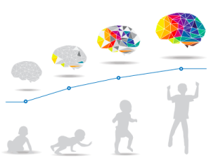
Some of the most dramatic physical change that occurs during this period is in the brain. At birth, the brain is about 25 percent of its adult weight, and this is not true for any other part of the body. By age 2, it is at 75 percent of its adult weight, at 95 percent by age 6, and at 100 percent by age 7 years.

Organization of high-level visual cortex in human infants”. Image retrieved from https://www.quantamagazine.org/infant-brains-reveal-how-the-mind-gets-built-20170110/.
We are born with most of the brain cells that we will ever have; that is, about 85 billion neurons whose function is to store and transmit information (Huttenlocher & Dabholkar, 1997). While most of the brain’s neurons are present at birth, they are not fully mature. During the next several years dendrites, or branching extensions that collect information from other neurons, will undergo a period of transient exuberance or temporary dramatic growth (exuberant because it is so rapid and transient because some of it is temporary). Because of this proliferation of dendrites, by age two a single neuron might have thousands of dendrites. Synaptogenesis, or the formation of connections between neurons, continues from the prenatal period forming thousands of new connections during infancy and toddlerhood. This period of rapid neural growth is referred to as synaptic blooming.
The blooming period of neural growth is then followed by a period of synaptic pruning, where neural connections are reduced thereby making those that are used much stronger. It is thought that pruning causes the brain to function more efficiently, allowing for mastery of more complex skills (Kolb & Whishaw, 2011). Experience will shape which of these connections are maintained and which of these are lost. Ultimately, about 40 percent of these connections will be lost (Webb et al., 2001). Blooming occurs during the first few years of life, and pruning continues through childhood and into adolescence in various areas of the brain.
Another major change occurring in the central nervous system is the development of myelin, a coating of fatty tissues around the axon of the neuron (Carlson, 2014). Myelin helps insulate the nerve cell and speed the rate of transmission of impulses from one cell to another. This enhances the building of neural pathways and improves coordination and control of movement and thought processes. The development of myelin continues into adolescence but is most dramatic during the first several years of life.
The infant brain grows very fast. At birth the brain is about 250 grams (half a pound) and by one year it is already 750 grams (Eliot, 1999). Comparing to adult size, the newborn brain is approximately 33% of adult size at birth, and in just 90 days, it is already at 55% of adult size (Holland et al., 2014). Most of the neural activity is occurring in the cortex or the thin outer covering of the brain involved in voluntary activity and thinking. The cortex is divided into two hemispheres, and each hemisphere is divided into four lobes, each separated by folds known as fissures. If we look at the cortex starting at the front of the brain and moving over the top, we see first the frontal lobe (behind the forehead), which is responsible primarily for thinking, planning, memory, and judgment. Following the frontal lobe is the parietal lobe, which extends from the middle to the back of the skull and which is responsible primarily for processing information about touch. Next is the occipital lobe, at the very back of the skull, which processes visual information. Finally, in front of the occipital lobe, between the ears, is the temporal lobe, which is responsible for hearing and language (Jarrett, 2015).
Watch It
This brief video describes some of the remarkable brain development that takes places in the first few years of life.
You can view the transcript for “How baby brains develop” here (opens in new window).
Although the brain grows rapidly during infancy, specific brain regions do not mature at the same rate. Primary motor areas develop earlier than primary sensory areas, and the prefrontal cortex, that is located behind the forehead, is the least developed (Giedd, 2015). As the prefrontal cortex matures, the child is increasingly able to regulate or control emotions, to plan activities, strategize, and have better judgment. This is not fully accomplished in infancy and toddlerhood, but continues throughout childhood, adolescence and into adulthood.
Lateralization is the process in which different functions become localized primarily on one side of the brain. For example, in most adults the left hemisphere is more active than the right during language production, while the reverse pattern is observed during tasks involving visuospatial abilities (Springer & Deutsch, 1993). This process develops over time, however, structural asymmetries between the hemispheres have been reported even in fetuses (Chi et al., 1997; Kasprian et al., 2011) and infants (Dubois et al., 2009).
Lastly, neuroplasticity refers to the brain’s ability to change, both physically and chemically, to enhance its adaptability to environmental change and compensate for injury. The control of some specific bodily functions, such as movement, vision, and hearing, is performed in specified areas of the cortex, and if these areas are damaged, the individual will likely lose the ability to perform the corresponding function. The brain’s neurons have a remarkable capacity to reorganize and extend themselves to carry out these particular functions in response to the needs of the organism, and to repair any damage. As a result, the brain constantly creates new neural communication routes and rewires existing ones. Both environmental experiences, such as stimulation and events within a person’s body, such as hormones and genes, affect the brain’s plasticity. So too does age. Adult brains demonstrate neuroplasticity, but they are influenced less extensively than those of infants (Kolb & Fantie, 1989; Kolb & Whishaw, 2011).
Watch It
How does all of this brain growth translate into cognitive abilities? We will discuss this in our next unit, but this video provides a nice overview of new research and some of the impressive abilities of newborns.
You can view the transcript for “Babies are Surprisingly Smart” here (opens in new window).
Try It
The Adolescent Brain
The brain undergoes dramatic changes during adolescence. Although it does not get larger, it matures by becoming more interconnected and specialized (Giedd, 2015). The myelination and development of connections between neurons continues. This results in an increase in the white matter of the brain that allows the adolescent to make significant improvements in their thinking and processing skills. Different brain areas become myelinated at different times. For example, the brain’s language areas undergo myelination during the first 13 years. Completed insulation of the axons consolidates these language skills but makes it more difficult to learn a second language. With greater myelination, however, comes diminished plasticity as a myelin coating inhibits the growth of new connections (Dobbs, 2012).
Even as the connections between neurons are strengthened, synaptic pruning occurs more than during childhood as the brain adapts to changes in the environment. This synaptic pruning causes the gray matter of the brain, or the cortex, to become thinner but more efficient (Dobbs, 2012). The corpus callosum, which connects the two hemispheres, continues to thicken allowing for stronger connections between brain areas. Additionally, the hippocampus becomes more strongly connected to the frontal lobes, allowing for greater integration of memory and experiences into our decision making.
The Teen Brain: 6 Things to Know
As you learn about brain development during adolescence, consider these six facts from the The National Institute of Mental Health:
Your brain does not keep getting bigger as you get older
For girls, the brain reaches its largest physical size around 11 years old and for boys, the brain reaches its largest physical size around age 14. Of course, this difference in age does not mean either boys or girls are smarter than one another!
But that doesn’t mean your brain is done maturing
For both boys and girls, although your brain may be as large as it will ever be, your brain doesn’t finish developing and maturing until your mid- to late-20s. The front part of the brain, called the prefrontal cortex, is one of the last brain regions to mature. It is the area responsible for planning, prioritizing and controlling impulses.
The teen brain is ready to learn and adapt
In a digital world that is constantly changing, the adolescent brain is well prepared to adapt to new technology—and is shaped in return by experience.
Many mental disorders appear during adolescence
All the big changes the brain is experiencing may explain why adolescence is the time when many mental disorders—such as schizophrenia, anxiety, depression, bipolar disorder, and eating disorders—emerge.
The teen brain is resilient
Although adolescence is a vulnerable time for the brain and for teenagers in general, most teens go on to become healthy adults. Some changes in the brain during this important phase of development actually may help protect against long-term mental disorders.
Teens need more sleep than children and adults
Although it may seem like teens are lazy, science shows that melatonin levels (or the “sleep hormone” levels) in the blood naturally rise later at night and fall later in the morning than in most children and adults. This may explain why many teens stay up late and struggle with getting up in the morning. Teens should get about 9-10 hours of sleep a night, but most teens don’t get enough sleep. A lack of sleep makes paying attention hard, increases impulsivity and may also increase irritability and depression.
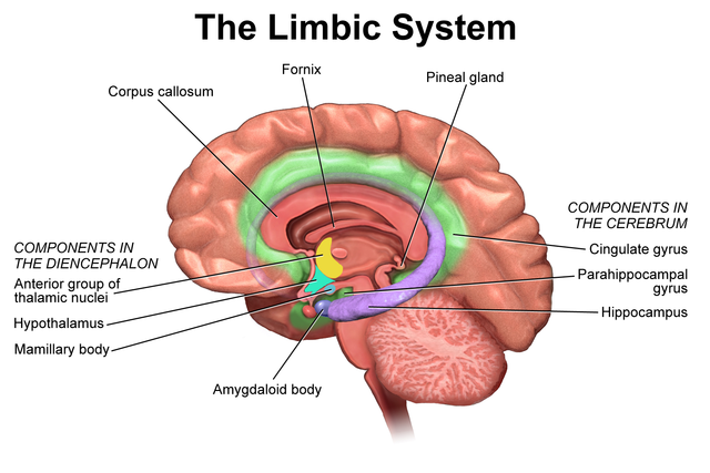
The limbic system, which regulates emotion and reward, is linked to the hormonal changes that occur at puberty. The limbic system is also related to novelty seeking and a shift toward interacting with peers. In contrast, the prefrontal cortex which is involved in the control of impulses, organization, planning, and making good decisions, does not fully develop until the mid-20s. According to Giedd (2015), an important outcome of the early development of the limbic system combined with the later development of the prefrontal cortex is the “mismatch” in timing between the two. The approximately ten years that separate the development of these two brain areas can result in increases in risky behavior, poor decision making, and weak emotional control for the adolescent. When puberty begins earlier, this mismatch lasts even longer.
Teens typically take more risks than adults and according to research it is because they weigh risks and rewards differently than adults do (Dobbs, 2012). The brain’s sensitivity to the neurotransmitter dopamine peaks during adolescence, and dopamine is involved in reward circuits, so adolescents may judge that the possible rewards outweigh the risks. Adolescents respond especially strongly to social rewards during activities, and they prefer the company of others their same age. Chein et al. (2011) found that peers sensitize brain regions associated with potential rewards. For example, adolescent drivers make more risky driving decisions when with friends to impress them, and teens are much more likely to commit crimes together in comparison to adults (30 and older) who commit them alone (Steinberg et al., 2018). In addition to dopamine, the adolescent brain is affected by oxytocin which facilitates bonding and makes social connections more rewarding. With both dopamine and oxytocin engaged, it is no wonder that adolescents seek peers and excitement in their lives that could actually end up endangering them.
Watch It
Watch the selected portion of this video to learn more about research related to brain changes and behavior during adolescence.
You can view the transcript for “The Teenage Brain Explained” here (opens in new window).
To learn more, watch this TED talk by Sarah-Jayne Blakemore: The mysterious workings of the adolescent brain about the latest adolescent brain research and more about how these changes in brain development also result in behavioral changes.
Because of all the changes that occur in the brain during adolescence, the chances for abnormal development, including the emergence of mental illness, also rise. In fact, 50% of all mental illnesses occur by the age 14 and 75% occur by age 24 (Giedd, 2015). Additionally, during this period of development the adolescent brain is especially vulnerable to damage from drug exposure. For example, repeated exposure to marijuana can affect cellular activity in the endocannabinoid system. Consequently, adolescents are more sensitive to the effects of repeated marijuana exposure (Weir, 2015).
However, researchers have also focused on the highly adaptive qualities of the adolescent brain which allow the adolescent to move away from the family towards the outside world (Dobbs, 2012; Giedd, 2015). Novelty seeking and risk taking can generate positive outcomes including meeting new people and seeking out new situations. Separating from the family and moving into new relationships and different experiences are actually quite adaptive– for adolescents and for society.
Link to Learning
Optional Reading:
Social cognitive development during adolescence.
Suparna Choudhury, Sarah-Jayne Blakemore, Tony Charman Social Cognitive and Affective Neuroscience, Volume 1, Issue 3, December 2006, Pages 165–174, https://doi.org/10.1093/scan/nsl024
Watch It
This video further explains and highlights some of the key developments in the brain during adolescence.
Key Takeaways
In sum, the adolescent years are a time of intense brain changes. Interestingly, two of the primary brain functions develop at different rates. Brain research indicates that the part of the brain that perceives rewards from risk, the limbic system, kicks into high gear in early adolescence. The part of the brain that controls impulses and engages in longer-term perspective, the frontal lobes, matures later. This may explain why teens in mid-adolescence take more risks than older teens. As the frontal lobes become more developed, two things happen. First, self-control develops as teens are better able to assess cause and effect. Second, more areas of the brain become involved in processing emotions, and teens become better at accurately interpreting others’ emotions.[1]
The Brain in Late Adulthood
Research has demonstrated that the brain loses 5% to 10% of its weight between 20 and 90 years of age (Fjell & Walhovd, 2010). This decrease in brain volume appears to be due to the shrinkage of neurons, decreases in the number of synapses, and increasingly shorter axon lengths. According to Garrett (2015), normal declines in cognitive ability throughout the lifespan are associated with brain changes, including reduced activity of genes involved in memory storage, synaptic pruning, plasticity, and glutamate and GABA (neurotransmitters) receptors.
There is also a loss in white matter connections between brain areas. Without myelin, neurons demonstrate slower conduction and impede each other’s actions. A loss of synapses occurs in specific brain areas, including the hippocampus (involved in memory) and the basal forebrain region. Older individuals also activate larger regions of their attentional and executive networks, located in the parietal and prefrontal cortex, when they perform complex tasks. This increased activation coincides with reduced performance on both executive tasks and tests of working memory when compared to that of younger people (Kolb & Whishaw, 2011).
Continued Neurogenesis
Researchers at the University of Chicago found that new neurons continue to form into old age. Tobin et al. (2019) examined post-mortem brain tissue of individuals between the ages of 79 and 99 (average age 90.6) and found evidence of neurogenesis in the hippocampus. Approximately 2000 neural progenitor cells and 150, 000 developing neurons were found per brain, although the number of developing neurons was lower in people with cognitive impairments or Alzheimer’s disease. Tobin et al. (2019) hypothesized that the lower levels of neurogenesis in the hippocampus were associated with symptoms of cognitive decline and reduced synaptic plasticity.
The brain in late adulthood also exhibits considerable plasticity, and through practice and training, the brain can be modified to compensate for any age-related changes (Erber & Szuchman, 2015). Park and Reuter-Lorenz (2009) proposed the Scaffolding Theory of Aging and Cognition which states that the brain adapts to neural atrophy (dying of brain cells) by building alternative connections, referred to as scaffolding. This scaffolding allows older brains to retain high levels of performance. Brain compensation is especially noted in the additional neural effort demonstrated by those individuals who are aging well. For example, older adults who performed just as well as younger adults on a memory task used both prefrontal areas, while only the right prefrontal cortex was used in younger participants (Cabeza et al., 2002). Consequently, this decrease in brain lateralization appears to assist older adults with their cognitive skills.

Neurocognitive Disorders
As the brain ages, there are several common disorders that results from changes in brain functioning that impact cognition and personality.
Dementia
Dementia is the umbrella category used to describe the general long-term and often gradual decrease in the ability to think and remember that affects a person’s daily functioning. The manual used to help classify and diagnose mental disorders, the Diagnostic and Statistical Manual of Mental Disorders, or DSM-5, classifies dementia as a “major neurocognitive disorder,” with milder symptoms classified as “mild cognitive impairment,” although the term dementia is still in common use. Dementia generally refers to severely impaired judgment, memory, or problem-solving ability. It can occur before old age and is not an inevitable development even among the very old. Common symptoms of dementia include emotional problems, difficulties with language, and a decrease in motivation. A person’s consciousness is usually not affected. Globally, dementia affected about 46 million people in 2015. About 10% of people develop the disorder at some point in their lives, and it becomes more common with age. About 3% of people between the ages of 65–74 have dementia, 19% between 75 and 84, and nearly half of those over 85 years of age. In 2015, dementia resulted in about 1.9 million deaths, up from 0.8 million in 1990. As more people are living longer, dementia is becoming more common in the population as a whole.
Dementia can be caused by numerous diseases and circumstances, all of which result in similar general symptoms of impaired judgment, etc. Alzheimer’s disease is the most common form of dementia and is incurable, but there are also nonorganic causes of dementia which can be prevented. Malnutrition, alcoholism, depression, and mixing medications can also result in symptoms of dementia. If these causes are properly identified, they can be treated. Cerebral vascular disease can also reduce cognitive functioning.
Alzheimer’s Disease
Alzheimer’s disease (AD), also referred to simply as Alzheimer’s, is the most common cause of dementia, accounting for 60-70% of its cases. Alzheimer’s is a progressive disease causing problems with memory, thinking, and behavior. Symptoms usually develop slowly and get worse over time, becoming severe enough to interfere with daily tasks.[2]
The most common early symptom is difficulty in remembering recent events. As the disease advances, symptoms can include problems with language, disorientation (including easily getting lost), mood swings, loss of motivation, not managing self-care, and behavioral issues. In the early stages, memory loss is mild, but with late-stage Alzheimer’s, individuals lose the ability to carry on a conversation and respond to their environment.

Alzheimer’s is the sixth leading cause of death in the United States. On average, a person with Alzheimer’s lives four to eight years after diagnosis but can live as long as 20 years, depending on other factors. Alzheimer’s is not a normal part of aging. The greatest known risk factor is increasing age, and the majority of people with Alzheimer’s are 65 and older. But Alzheimer’s is not just a disease of old age. Approximately 200,000 Americans under the age of 65 have younger-onset Alzheimer’s disease (also known as early-onset Alzheimer’s).[3]
The cause of Alzheimer’s disease is poorly understood. About 70% of the risk is believed to be inherited from a person’s parents with many genes usually involved. Other risk factors include a history of head injuries, depression, and hypertension. The disease process is associated with plaques and neurofibrillary tangles in the brain. A probable diagnosis is based on the history of the illness and cognitive testing with medical imaging and blood tests to rule out other possible causes. Initial symptoms are often mistaken for normal aging, but examination of brain tissue, specifically of structures called plaques and tangles, is needed for a definite diagnosis. Though qualified physicians can be up to 90% certain of a correct diagnosis of Alzheimer’s, currently, the only way to make a 100% definitive diagnosis is by performing an autopsy of the person and examining the brain tissue. In 2015, there were approximately 29.8 million people worldwide with AD. In developed countries, AD is one of the most financially costly diseases.
Watch It
This Ted-Ed video explains some of the history and biological diagnosis of Alzheimer’s.
You can view the transcript for “What is Alzheimer’s disease? – Ivan Seah Yu Jun” here (opens in new window).
Link to Learning
Samuel Cohen researches Alzheimer’s disease and other neurodegenerative disorders. Listen to Cohen’s TED Talk on Alzheimer’s disease to learn more.
Try It
Delirium
Delirium, also known as acute confusional state, is an organically caused decline from a previous baseline level of mental function that develops over a short period of time, typically hours to days. It is more common in older adults, but can easily be confused with a number of psychiatric disorders or chronic organic brain syndromes because of many overlapping signs and symptoms in common with dementia, depression, psychosis, etc. Delirium may manifest from a baseline of existing mental illness, a baseline intellectual development disorder, or dementia, without being due to any of these problems.
Delirium is a syndrome encompassing disturbances in attention, consciousness, and cognition. It may also involve other neurological deficits, such as psychomotor disturbances (e.g. hyperactive, hypoactive, or mixed), impaired sleep-wake cycle, emotional disturbances, and perceptual disturbances (e.g. hallucinations and delusions), although these features are not required for diagnosis. Among older adults, delirium occurs in 15-53% of post-surgical patients, 70-87% of those in the ICU, and up to 60% of those in nursing homes or post-acute care settings. Among those requiring critical care, delirium is a risk factor for death within the next year.
Healthy Brain Functioning
In longitudinal studies, Cheng (2016) found that both physical activity and stimulating cognitive activity resulted in significant reductions in the risk of neurocognitive disorders. Physical activity, especially aerobic exercise, is associated with less age-related gray and white matter loss, as well and diminished neurotoxins in the brain.
Overall, physical activity preserves the integrity of neurons and brain volume. Cognitive training improves the efficiency of the prefrontal cortex and executive functions, such as working memory, and strengthens the plasticity of neural circuits. Both activities support cognitive reserve, or “the structural and dynamic capacities of the brain that buffer against atrophies and lesions” (Cheng, 2016, p. 85). Although it is optimal to begin physical and cognitive activities earlier in life, it is never too late to start these programs to improve one’s cognitive health, even in late adulthood.
Can we improve brain functioning?
Many training programs have been created to improve brain functioning. ACTIVE (Advanced Cognitive Training for Independent and Vital Elderly), a study conducted between 1999 and 2001 in which 2,802 individuals age 65 to 94, suggests that the answer is “yes”. These racially diverse participants received 10 group training sessions and 4 follow up sessions to work on tasks of memory, reasoning, and speed of processing. These mental workouts improved cognitive functioning even 5 years later. Many of the participants believed that this improvement could be seen in everyday tasks as well (Tennstedt et al., 2006).
However, programs for the elderly on memory, reading, and processing speed training demonstrate that there is improvement on the specific tasks trained, but there is no generalization to other abilities (Jarrett, 2015). Further, these programs have not been shown to delay or slow the progression of Alzheimer’s disease. Although these programs are not harmful, “physical exercise, learning new skills, and socializing remain the most effective ways to train your brain” (p. 207). These activities appear to build a reserve to minimize the effects of primary aging of the brain.
References (Click to expand)
Cabeza, R., Anderson, N. D., Locantore, J. K., & McIntosh, A. R. (2002). Aging gracefully: Compensatory brain activity in highperforming older adults. NeuroImage, 17, 1394-1402.
Carlson, N. (2014). Foundations of behavioral neuroscience (9th ed.). Pearson.
Chein, J., Albert, D., O’Brien, L., Uckert, K., & Steinberg, L. (2011). Peers increase adolescent risk taking by enhancing activity in the brain’s reward circuitry. Developmental Science, 14(2), F1-F10. doi: 10.1111/j.1467-7687.2010.01035.x
Cheng, S. (2016). Cognitive reserve and the prevention of dementia: The role of physical and cognitive activities. Current Psychiatry Reports, 18(9), 85.
Chi, J. G., Dooling, E. C., & Gilles, F. H. (1977). Left-right asymmetries of the temporal speech areas of the human fetus. Archives of Neurology, 34, 346–8.
Dubois, J., Hertz-Pannier, L., Cachia, A., Mangin, J. F., Le Bihan, D., & Dehaene-Lambertz, G. (2009). Structural asymmetries in the infant language and sensori-motor networks. Cerebral Cortex, 19, 414–423.
Dobbs, D. (2012). Beautiful brains. National Geographic, 220(4), 36.
Eliot, L. (1999). What’s going on in there? Bantam.
Erber, J. T., & Szuchman, L. T. (2015). Great myths of aging. John Wiley & Sons.
Fjell, A. M., & Walhovd, K. B. (2010). Structural brain changes in aging: Courses, causes, and cognitive consequences. Reviews in the Neurosciences, 21, 187-222.
Garrett, B. (2015). Brain and behavior (4th ed.) Sage.
Giedd, J. N. (2015). The amazing teen brain. Scientific American, 312(6), 32-37.
Holland, D., Chang, L., Ernst, T., Curan, M…….. Dale, A. (2014). Structural growth trajectories and rates of change in the first 3 months of infant brain development. JAMA Neurology, 71(10), 1266-1274.
Huttenlocher, P. R., & Dabholkar, A. S. (1997). Regional differences in synaptogenesis in human cerebral cortex. Journal of comparative Neurology, 387(2), 167-178.
Jarrett, C. (2015). Great myths of the brain. Wiley
Kasprian, G., Langs, G., Brugger, P. C., Bittner, M., Weber, M., Arantes, M., & Prayer, D. (2011). The prenatal origin of hemispheric asymmetry: an in utero neuroimaging study. Cerebral Cortex, 21, 1076–1083.
Kolb, B., & Fantie, B. (1989). Development of the child’s brain and behavior. In C. R. Reynolds & E. Fletcher-Janzen (Eds.), Handbook of clinical child neuropsychology (pp. 17–39). Plenum Press.
Kolb, B. & Whishaw, I. Q. (2011). An introduction to brain and behavior (3rd ed.). Worth Publishers.
Park, D. C., & Reuter-Lorenz, P. (2009). The adaptive brain: Aging and neurocognitive scaffolding. Annual Review of Psychology, 60, 173-196.
Springer, S. P. & Deutsch, G. (1993). Left brain, right brain (4th ed.). W. H. Freeman.
Steinberg, L. (2008) A social neuroscience perspective on adolescent risk-taking. Developmental Review, 28:78-106.
Steinberg, L., Icenogle, G., Shulman, E.P., et al. (2018). Around the world, adolescence is a time of heightened sensation seeking and immature self-regulation. Developmental Science, 21, e12532. https://doi.org/10.1111/desc.12532
Tennstedt, S., Morris, J., Unverzagt, F., Rebok, G., Willis, S., Ball, K., & Marsiske, M. (2006). ACTIVE: Advanced Cognitive Training for Independent and Vital Elderly Clinical Trial. Clinical Trials Database and Worldwide Listings. Retrieved from http://www.clinicaltrialssearch.org/active-advanced-cognitive-training-for-independent-and-vital-elderlynct00298558.html
Tobin, M., K., Musaraca, K., Disouky, A., Shetti, A., Bheri, A. Honer, W. G.,…Lazarov, O. (2019). Human hippocampal neurogenesis persists in aged adults and Alzheimer’s disease patients. Cell Stem Cell, 24(6), 974-982.
Webb, S. J., Monk, C. S., & Nelson, C. A. (2001). Mechanisms of postnatal neurobiological development: Implications for human development. Developmental Neuropsychology, 19, 147-171.
Weir, K. (2015). Marijuana and the developing brain. Monitor on Psychology, 46(10), 49-52
Licenses & Attributions (Click to expand)
CC Licensed Content
- “Physical Growth & Brain Development.” Authored by: Tera Jones for Lumen Learning. Provided by: Lumen Learning. Located at: https://courses.lumenlearning.com/wm-lifespandevelopment/chapter/physical-development/. License: CC BY: Attribution
- Psyc 200 Lifespan Psychology. Authored by: Laura Overstreet. Located at: http://opencourselibrary.org/econ-201/. License: CC BY: Attribution
- Childhood: Physical and Cognitive Development image and supportive text. Provided by: Lumen Learning. Located at: https://courses.lumenlearning.com/waymaker-psychology/chapter/reading-childhood/. Project: Introduction to Psychology. License: CC BY: Attribution
- “Lifespan Development: A Psychological Perspective, Second Edition” by Martha Lally and Suzanne Valentine-French is licensed under a CC-BY-NC-SA-3.0
- “Puberty & Cognition” by Ellen Skinner, Brandy Brennan, & Dan Grimes is licensed under a CC-BY-NC-SA-3.0.
- Lifespan Development by Lumen Learning is licensed under a Creative Commons Attribution 4.0 International License
- “Cognitive Function in Late Adulthod” Authored by: Sonja Ann Miller for Lumen Learning. Provided by: Lumen Learning. License: CC BY: Attribution
- Alzheimer’s Disease. Provided by: Wikipedia. Located at: https://en.wikipedia.org/wiki/Alzheimer%27s_disease. License: CC BY-SA: Attribution-ShareAlike
- Delirium. Provided by: Wikipedia. Located at: https://en.wikipedia.org/wiki/Delirium. License: CC BY-SA: Attribution-ShareAlike
- Brain Development During Adolescence modification, adaptation, and original content. Authored by: Tera Jones for Lumen Learning. Provided by: Lumen Learning. Located at: https://courses.lumenlearning.com/wm-lifespandevelopment/chapter/brain-development-during-adolescence/. License: CC BY-SA: Attribution-ShareAlike
Media Attributions
- LimbicSystem © Bruce Blaus is licensed under a CC BY (Attribution) license
- exercisebrain © Willian Murphy is licensed under a CC BY-SA (Attribution ShareAlike) license
- Alzheimer’s disease brain comparison. Authored by: Garrando. Provided by: Wikipedia. Located at: https://en.wikipedia.org/wiki/Dementia#/media/File:Alzheimer%27s_disease_brain_comparison.jpg. License: Public Domain: No Known Copyright
All Rights Reserved Content
- How Baby Brains Develop. Provided by: CNN. Located at: https://www.youtube.com/watch?v=R0fiu2S0_3M. License: Other. License Terms: Standard YouTube License
- Baby MRI image. Provided by: Quanta Magazine. Located at: https://www.quantamagazine.org/infant-brains-reveal-how-the-mind-gets-built-20170110/. License: All Rights Reserved
- Babies are surprisingly smart. Authored by: SciShow Psych. Located at: https://www.youtube.com/watch?v=nM-GhFzX8yQ. License: Other. License Terms: Standard YouTube License
- What is Alzheimer’s. Provided by: Ted-Ed. Located at: https://www.youtube.com/watch?v=yJXTXN4xrI8. License: All Rights Reserved
- The Teenage Brain Explained. Provided by: SciShow. Located at: https://www.youtube.com/watch?v=hiduiTq1ei8&feature=youtu.be. License: Other. License Terms: Standard YouTube License
- Brain changes during adolescence | Behavior | MCAT. Provided by: Khan Academy. Located at: https://www.youtube.com/watch?v=5Fa8U6BkhNo&feature=youtu.be. License: Other. License Terms: Standard YouTube License
- Steinberg, L. (2008) A social neuroscience perspective on adolescent risk-taking. Developmental Review, 28:78-106. ↵
- What is Alzheimer's? Alzheimer's Association. Retrieved from https://www.alz.org/alzheimers-dementia/what-is-alzheimers ↵
- What is Alzheimer's? Alzheimer's Association. Retrieved from https://www.alz.org/alzheimers-dementia/what-is-alzheimers. ↵
